高血壓 高尿酸 慢性腎病 胰島素 https://2019medicinenote.blogspot.com/2019/12/blog-post_57.html . 糖尿病相關筆記~目錄 https://2019medicinenote.blogspot.com/2020/01/blog-post_4.html

2023年12月28日 星期四

急性鼻竇炎 -台灣家庭醫學醫學會及uptodate . StatPearls [Internet].

2024-08-09 10:34AM 
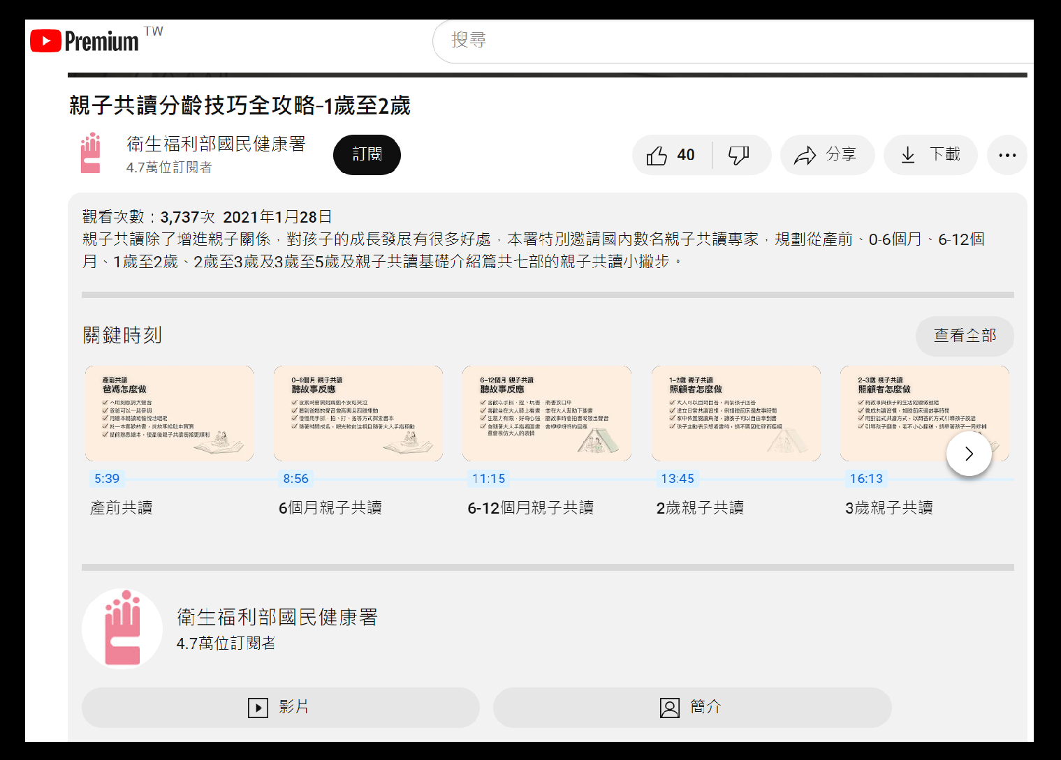
剛看到ENT醫師寫的文章. 提到急性鼻竇炎標準治療是抗生素.
順手查了一篇2023年8月的paper. 關於急性鼻竇炎定義. 四周內屬於急性. 
而要診斷急性鼻竇炎. 症狀通常持續 10 天以上. 
因此. 一般感冒初期一周. 不開抗生素是合理的. 


google中文翻譯如下(原文在下面, 僅節錄片段)
病毒性鼻竇炎(VRS)
急性細菌性鼻竇炎 (ABRS) 
急性侵襲性真菌性鼻竇炎(IFS)
過敏性真菌性鼻竇炎(AFS)

鼻竇炎可分為以下幾類(更多基於共識而非實證研究)
急性 - 症狀持續少於 4 週
亞急性 - 症狀持續 4 至 12 週
慢性 - 症狀持續超過 12 週
復發 - 四次發作,持續時間不到 4 週,兩次發作之間症狀完全緩解

急性鼻竇炎是一種臨床診斷。臨床醫生最常需要區分 VRS 和 ABRS,這對於確保負責任地使用抗生素至關重要。需要闡明青黴素不敏感肺炎鏈球菌的當地抗藥性模式和盛行率。

成人鼻竇炎的常規診斷標準是患者至少有兩種主要症狀或一種主要症狀加上兩種或多種輕微症狀。兒童的標準相似,只是更注重鼻分泌物(而非鼻塞)。

主要症狀

  • 前鼻流膿性分泌物
  • 後鼻涕膿性或變色
  • 鼻塞或阻塞
  • 臉部充血或飽滿
  • 臉部疼痛或壓力
  • 嗅覺減退或嗅覺喪失
  • 發燒(僅適用於急性鼻竇炎) 

輕微症狀:

  • 頭痛
  • 耳朵疼痛或有壓力或飽脹感
  • 口臭
  • 牙齒疼痛
  • 咳嗽
  • 發燒(亞急性或慢性鼻竇炎)
  • 疲勞

可以使用以下臨床指導將 ABRS 與 VRS 區分開來

  • 症狀持續十天以上
  • 發病時持續 3 至 4 天高燒(超過 39°C 或 102°F),伴隨膿性鼻涕或臉部疼痛
  • 前十天內症狀加倍惡化

一般不需要進行常規實驗室評估。囊性纖維化、纖毛功能障礙或免疫缺陷的評估是頑固性、復發性或慢性鼻竇炎的考慮因素。 有證據顯示 ESR 和 CRP 升高可能與細菌感染有關。

大於或等於 10 CFU/mL 的內視鏡抽吸培養物被視為黃金標準。然而,這對於診斷來說並不是必需的,並且對於絕大多數 ABRS 病例也沒有這樣做。鼻和鼻咽培養物的實用性較低,因為它們與內視鏡抽吸物的相關性較差。轉診內視鏡抽吸對於難治性病例或多種抗生素過敏的患者可能有用。  

除非臨床上擔心併發症或替代診斷,否則沒有必要急性鼻竇炎進行影像學檢查。竇平片通常不能用於檢測發炎。它們可能會顯示空氣-液體水平。然而,這無助於區分病毒與細菌病因。如果懷疑有併發症或替代診斷,或患者反覆出現急性感染,則應進行鼻竇 CT 影像,以評估骨骼、軟組織、牙齒或其他解剖異常或是否有慢性鼻竇炎。這些應該在適當的治療過程結束時獲得。鼻竇 CT 可顯示氣液水平、混濁和發炎。鼻竇黏膜增厚超過 5 毫米則表示有發炎。 它還可以有效評估骨侵蝕或破壞。然而,這些發現也無助於區分病毒與細菌病因。 MRI 比鼻竇 CT 提供更多細節,可以評估軟組織或幫助闡明腫瘤。因此,MRI 可能有助於確定眼眶或顱內擴展等病例的併發症程度。


Evaluation
Acute rhinosinusitis is a clinical diagnosis. The clinician most commonly needs to distinguish between VRS and ABRS, which is crucial to ensure the responsible usage of antibiotics. Local resistance patterns and prevalence of penicillin non-susceptible S. pneumoniae requires elucidation.

Conventional diagnostic criteria for rhinosinusitis in adults is the patient having at least two major or one major plus two or more minor symptoms. The criteria in children are similar except that there is more of an emphasis on nasal discharge (rather than nasal obstruction).Major symptoms:Purulent anterior nasal discharge
Purulent or discolored posterior nasal discharge
Nasal congestion or obstruction
Facial congestion or fullness
Facial pain or pressure
Hyposmia or anosmia
Fever (for acute sinusitis only)

Minor symptoms:Headache
Ear pain or pressure or fullness
Halitosis
Dental pain
Cough
Fever (for subacute or chronic sinusitis)
Fatigue

ABRS can be differentiated from VRS using the following clinical guidance[4][6][1][3]:Duration of symptoms for more than ten days
High fever (over 39 C or 102 F) with purulent nasal discharge or facial pain that last for 3 to 4 consecutive days at the beginning of the illness
Double worsening of symptoms within the first ten days

Routine laboratory assessment is generally not necessary. Evaluation for cystic fibrosis, ciliary dysfunction, or immunodeficiency are considerations for intractable, recurrent, or chronic rhinosinusitis. There is some evidence that an elevated ESR and CRP may be associated with a bacterial infection.

The culture of endoscopic aspirates with greater than or equal to 10 CFU/mL is considered the gold standard. However, this is not necessary for diagnosis and not done for the vast majority of cases of ABRS. Nasal and nasopharyngeal cultures are of low utility due to their poor correlation with endoscopic aspirates. Referral for endoscopic aspiration can be useful for refractory cases or patients with multiple antibiotic allergies.

Imaging for acute sinusitis is not necessary unless there is a clinical concern for a complication or alternative diagnosis. Sinus plain films are generally not useful in detecting inflammation. They may show air-fluid levels. However, this does not help differentiate viral versus bacterial etiologies. If a complication or alternative diagnosis is suspected, or if the patient has recurrent acute infections, then sinus CT imaging should be obtained to assess for bone, soft tissue, dental, or other anatomical abnormalities or the presence of chronic sinusitis. These should be obtained at the end of an appropriate treatment course. Sinus CT may show air-fluid levels, opacification, and inflammation. A thickened sinus mucosa over 5 mm is indicative of inflammation. It can also effectively assess bony erosion or destruction. However, these findings are also not helpful in differentiating viral versus bacterial etiologies. MRI offers more detail than sinus CT to evaluate soft tissue or help elucidate a tumor. Thus, MRI may be helpful to determine the extent of complications in cases such as an orbital or intracranial extension.

2024-05-14 11:45AM
參考資料: 成人急性細菌性鼻竇炎治療指引檔案下載
這篇是刊登於家庭醫學與基層醫療第二十八卷第5期(102 年 5 月 25 日)
雖資料有點陳舊., 不過需修改的地方不多.
(家庭醫學與基層醫療期刊總目錄)


參考資料 Acute sinusitis and rhinosinusitis in adults: Clinical manifestations and diagnosis uptodate
2023-12-29 10:02AM
病毒性鼻竇炎. 症狀通常小於10天.
Acute viral rhinosinusitis — Acute viral rhinosinusitis (AVRS) is diagnosed clinically when patients have <10 days of symptoms consistent with ARS that are not worsening [2].

細菌性鼻竇炎.
Acute bacterial rhinosinusitis — We use the following criteria to diagnose acute bacterial rhinosinusitis (ABRS), which are derived from the American Academy of Otolaryngology-Head and Neck Surgery guidelines and the Infectious Diseases Society of America guidelines [2,16]:


診斷標準(符合其中一項)
1. 症狀持續超過 10 天沒有改善
2. 感冒症狀持續超過十天. 期間症狀改善, 第五六天後又變嚴重.
●Persistent symptoms or signs of ARS lasting 10 or more days without evidence of clinical improvement or

●A biphasic pattern of illness, typically extending over a 10-day period, characterized by signs and symptoms of ARS that initially start to improve but then worsen approximately five to six days later ("double worsening").

嚴重感染症狀. 持續性高燒超過 39°C. 膿狀鼻涕. 顏面疼痛 3-4 天以上
但並非症狀嚴重就需要吃抗生素
The onset of severe symptoms or signs of severe illness (eg, high fever [>39°C or 102°F], purulent nasal discharge, facial pain) for at least three to four consecutive days at the beginning of illness supports the diagnosis of ABRS. However, severity of illness alone is not sufficient criteria for starting antibiotics.

並非每個患者都需要做影像檢查. 鼻腔細菌培養, 鼻竇抽吸,
當懷疑患者發生併發症才需要做.
Imaging, nasal cultures, sinus aspirates, and other microbiologic testing are not indicated for patients with clinically diagnosed uncomplicated AVRS or ABRS [2,16]. These tests are reserved for patients with suspected complications.

Other guidelines use varying criteria to diagnose ABRS. These criteria may be based on specific symptoms or duration of illness [37,38].

2023年12月21日 星期四

[疾管署2023-09-01 ]流感疫苗副作用

2023-12-22 
路徑: 疾管署首頁 >>預防接種 >>疫苗資訊 >>疫苗區Q&A 季節性流感疫苗Q&A 疫苗安全及接種篇

Q27.流感疫苗安全嗎?會有什麼副作用?
今年政府採購4廠牌疫苗皆符合我國衛生福利部食品藥物管理署查驗登記規定,且經其核准使用/進口,安全無虞。

疫苗與其他任何藥品一樣有可能造成副作用,包括接種後可能會有注射部位疼痛、紅腫,少數的人則會有全身性的輕微反應,如:發燒、頭痛、肌肉酸痛、噁心、皮膚搔癢、蕁麻疹或紅疹等,一般會在發生後1-2天內自然恢復。

嚴重的副作用,如立即型過敏反應,甚至過敏性休克等不適情況(臨床表現包括呼吸困難、聲音沙啞、氣喘、眼睛或嘴唇腫脹、頭昏、心跳加速等),發生機率非常低,若不幸發生,通常於接種後幾分鐘至幾小時內即出現症狀。

其他曾被零星報告過之不良事件包括神經系統症狀(如:臂神經叢炎、顏面神經麻痺、熱痙攣、腦脊髓炎、以對稱性神經麻痺為表現的Guillain-Barré症候群等)和血液系統症狀(如:暫時性血小板低下,臨床表現包括皮膚出現紫斑或出血點、出血時不易止血等)。

除了1976年豬流感疫苗、2009年H1N1新型流感疫苗與部分季節性流感疫苗經流行病學研究證實與Guillain-Barré症候群可能相關外,少有確切統計數據證明其他不良事件與接種流感疫苗有關。

最後更新日期2023/9/1


2023年12月13日 星期三

B肝帶原患者肝指數上升評估

2023-12-14 15:10
B肝帶原患者. 肝指數上升需檢測病毒量. 如果少於 2000 iu/mL 需考慮其他原因

肝發炎指數異常!到底要不要緊?
諮詢╱徐士哲(臺大肝膽胃腸科主治醫師)楊培銘(臺大醫學院名譽教授)撰稿╱黃靜宜

即使有B肝帶原,肝發炎指數又異常,也不能斷定就是B肝引起,這時會加驗血中B肝病毒量。如果少於2000 IU/ml,甚至測不到病毒量,就要考慮其他可能性。

C肝治療

2023-12-13
C肝感染之後
7-8 成會變成慢性肝炎.
經過 20-30 年. 有 2-3 成的患者會進展到肝硬化
C肝合併肝硬化. 每年5%會罹患肝癌

C肝病毒抗體陽性. 代表曾經被感染過. 這樣的患者. 約 42~88% 會呈現 HCV RNA 陰性. 亦即體內已經沒有 C肝病毒
參考資料: 國家消除C肝政策綱領 2018~2025 (2019年5月出版)


[疾管署]
急性病毒性C型肝炎防治工作手冊

 附件: 急性病毒性C型肝炎工作手冊.pdf(2019年1月修訂)


[疾管署]急性病毒性C型肝炎核心衛教_1080830修.pdf

C型肝炎病毒急性感染後
約20-30%患者有臨床症狀(發燒、疲倦、厭食、隱約腹部不適、噁心、嘔吐或黃疸等)
 約70%-80%會演變成慢性肝炎,在慢性C型肝炎患者中,約5-20%於20-30年間可能演變為肝硬化,約1-5%死於慢性肝炎的併發症。
 目前無C型肝炎疫苗。

Hepatitis C and hepatocellular carcinoma
Among patients with hepatitis C and cirrhosis, the annual incidence rate of HCC ranges between 1-8%, being higher in Japan (4-8%) intermediate in Italy (2-4%) and lower in USA (1.4%).

[2012-04-15]C肝者罹肝癌風險 6指標預知
在台灣約有40~60萬人是C型肝炎患者。感染C型肝炎後,約七~八成的人會變成慢性C型肝炎;這些患者經過二十、三十年後,有二~三成演變成肝硬化,其中每年約有百分之五的人,會併發肝癌,因此C型肝炎病毒可說是僅次於B型肝炎的台灣肝病第二號殺手。


2020年3月1日開始. 使用健保開C肝治療藥物不設限.
病人只要確認感染慢性C肝,不論有無肝纖維化,均可成為給藥對象
參考資料: 好心肝雜誌(2020-04-15)

若病患知道自己有C肝. 但C肝抗體篩檢報告超過三年.
1. 第一次門診. 重新檢驗是否仍C肝抗體陽性. 如果仍C肝抗體陽性. 再做 RNC定量
2. 第二次門診. 因為治療前的抽血項目有 PT. 檢體不能放太久 所以需等C肝抗體和RNA>0 再來門診開立治療前的14項檢驗
3. 第三次門診開28天藥物. 當HCV基因型檢查出來之後. 聯繫病患. 再次確認病患願意吃藥治療. 聯繫廠商送藥(每周一). 確定藥品到達當天. 請病患掛門診開藥 順便開28天候抽血單.
4. 第4次門診, 開28天 藥物. 順便開下次檢驗單. 約病患回來抽血 5. 第5次門診. 治療結束後 12 周. 至門診開抽血單. 檢驗血中是否仍有HCV 病毒. 完成C肝療程(陽性機率<1/4000)

藥物使用. 以艾百樂(Maviret)為例. 每次三顆. 每天一次. 總共吃 56 天.

整個療程包括治療前抽血. 治療結束後 12 周抽血. 前後需要五個月.

其他相關資料
健保署新聞稿[110-11-29] 健保開放C肝全口服新藥處方不限科別 助攻C肝消除
yahoo新聞 C 型肝炎全口服新藥健保給付執行計畫


2023年12月6日 星期三

軟組織受傷使用冰敷已經過時?

2023-12-06 19:08
先回顧一下歷史. 冰敷是在 1978年由運動醫學醫師Gabe Mirkin提出的. 但在 2015 年 Gabe Mirkin 推翻了自己的建議.這段歷史可以參考下面兩個連結
 
參考資料1 . 下面中文是用google自動翻譯


參考資料2. 下面中文是用google自動翻譯. 裡面提到了Hyperbaric gaseous cryotherapy高壓氣體冷療法. 蠻有趣的. 順便將paper裡面表格附上.  
The R.I.C.E Protocol is a MYTH: A Review and Recommendations

摘要: 
1962 年 5 月 23 日,12 歲的埃弗雷特(埃迪)諾爾斯在馬薩諸塞州薩默維爾跳上一列貨運列車,導致他的手臂與身體完全分離。這名小男孩被送往馬薩諸塞州綜合醫院 (MGH),年輕的住院醫師羅納德·A·馬爾特 (Ronald A. Malt) 醫生試圖挽救艾迪的斷肢。儘管醫學文獻中從未記錄過重大肢體的成功再接,但 Malt 博士和 12 名醫生組成的團隊進行了歷史上首次成功的肢體再接。
On May 23, 1962, twelve- year- old Everett (Eddie) Knowles jumped on to a freight train in Somerville, MA resulting in his arm being completely severed from his body.  The young boy was taken to Massachusetts General Hospital (MGH) where Dr. Ronald A. Malt, a young chief resident, attempted to save Eddie’s detached limb.   Despite the fact that there had never been a successful reattachment of a major limb recorded in medical literature, Dr. Malt and a team of twelve doctors performed the first successful limb reattachment in history.
該行動的成功很快就成為全球現象。新聞播音員蜂擁而至,聚集在醫生團隊中,以獲取有關奇蹟般的肢體再接術的基本事實,該手術被譽為醫學史上最具有里程碑意義的手術之一。然而,手術的基本事實相當複雜,一般大眾無法理解。相反,記者將注意力集中在故事中引起讀者興趣的方面。因此,應用冰來保存切斷的組織成為故事的主要焦點。(21、33、36)
The operation’s success quickly became a global phenomenon.  Newscasters swarmed the team of doctors to obtain essential facts about the miracle limb reattachment touted as one of the most monumental operations in medical history.  However, the essential facts about the surgery were rather complicated and would not be understood by the general public.  Instead, reporters focused on the aspects of the story that would be intriguing to the reader.  As a result, the application of ice to preserve the severed tissue became the main focus of the story. (21, 33, 36)

在 1962 年 5 月 23 日事件之前,使用冰塊治療損傷從未成為醫療方案的一部分,而利用冰塊保存組織的想法很快就被全球各地的報紙報道。隨後,隨著沒有直接參與手術的個人不斷重述這個故事,事實開始改變。最終,大眾很快就接受了這樣的觀念:任何傷害都應該用冰敷來治療,無論其嚴重程度或如何發生 (36)。
The use of ice to treat injuries was never part of medical protocol prior to the events of May 23, 1962 and the notion to utilize ice for tissue preservation was quickly published by newspapers around the globe.  Subsequently, as the story was continuously retold by individuals not directly involved in the surgery, facts began to change.  Eventually, the general public was quickly accepting the notion that any injury should be treated with the application of ice, regardless of its severity or how it occurred (36).

1978年,Gabe Mirkin博士出版了《運動醫學書》,並創造了縮寫詞「RICE」(Rest、Ice、Compression、Elevation)來代表四種治療急性運動傷害的活動。四十多年來,RICE 協議已在學術課程和公眾認知中根深蒂固。然而,2013年,RICE在其著作《 Iced!》中受到了Gary Reinl的挑戰。虛幻的治療選擇。”  Reinl 引用了大量研究和解剖學資源來支持這一觀點,即在用冰緊緊包裹(壓縮)受傷部位的同時,休息並不能有效加速恢復過程,還可能導致受影響組織進一步受損。
In 1978, Dr. Gabe Mirkin released “Sportsmedicine Book” and coined the acronym “RICE” (Rest, Ice, Compression, and Elevation) to represent the four activities for treating acute athletic injuries.  The RICE protocol has been ingrained in academic curriculum as well as in public perception for over four decades.  In 2013, however, RICE was challenged by Gary Reinl in his book “Iced! The Illusionary Treatment Option.”  Reinl cited numerous studies and anatomical resources in support of the notion that resting an injury, while wrapping it tightly (compression) with ice, is ineffective in accelerating the recovery process and could also result in further damage to the affected tissues.

Reinl 的書發布後,Mirkin 在其個人網站 (31) 的 2015 年出版物中公開放棄了他對 RICE 協議的最初立場。米爾金甚至為萊因爾的第二版《 Ice d!》寫了序言。幻覺治療方案”,並對他所創造的方案提出了修改意見;
Following the release of Reinl’s book, Mirkin publicly recanted his original position on the RICE protocol in a 2015 publication on his personal website (31).  Mirkin even wrote the foreword to Reinl’s second edition of “Iced! The Illusionary Treatment Option”, and offered his revised opinion on the protocol he created;

隨後的研究表明,休息和冰敷實際上會延遲恢復。溫和的運動有助於組織更快癒合,而冷敷則可以抑制啟動並加速恢復的免疫反應。冰敷確實有助於抑制疼痛,但運動員通常更感興趣的是盡快返回比賽場地。因此,如今,RICE 並不是急性運動傷害的首選治療方法 (36)。
Subsequent research shows that rest and ice can actually delay recovery.    Mild    movement helps tissue to heal faster, and the application of cold suppresses the immune responses that start and hasten recovery. Icing does help suppress     pain, but athletes are usually far more interested in returning as quickly as possible to the playing field. So, today, RICE is not the preferred treatment for an acute athletic injury (36).

根據現有證據,唯一合理的結論是,使用 RICE 技術來加速恢復過程顯然是一個神話。2015 年,米爾金博士公開放棄了他 1978 年的最初立場,其有效性明顯受到損害。
Based upon the available evidence, the only plausible conclusion is that the use of the RICE technique to accelerate the recovery process is unequivocally a myth.  Its validity was unequivocally compromised in 2015 when Dr. Mirkin publicly recanted his original position from 1978.

有大量科學證據表明,有一些行之有效的方法可以加速肌肉、韌帶和肌腱損傷的癒合,但不包括長時間休息並結合冰敷、壓迫和抬高。為了揭穿 RICE 神話,謹慎的做法是探索對損傷的生理反應以及冰敷、壓迫、抬高和不活動對這些過程的影響。最終的結論是,有更優化的技術來加速恢復過程,其中不包括同時對受影響區域進行壓縮和局部冷卻(冰)的一段不活動期。
There is an abundance of scientific evidence purporting proven methods to accelerate the healing of muscle, ligament, and tendon injuries that do not include extended periods of rest used in conjunction with ice, compression, and elevation.  To debunk the RICE myth, it is prudent to explore the physiological responses to injury and the effect ice, compression, elevation and inactivity have on those processes.  The ultimate conclusion is that there are more optimal techniques to accelerate the recovery process that do not include a period of inactivity in which compression and topical cooling (ice) is simultaneously applied to the affected area.

Hyperbaric gaseous cryotherapy 表格的中文翻譯同樣是直接使用google翻譯. 


2023年11月30日 星期四

使用高濃度生理食鹽水校正低血鈉(網路計算機)

2023-12-01 14:23
網路上有很方便的計算器. 
HYPERTONIC SALINE 3% AND 0.9NS INFUSION RATE CALC

舉例. 70歲男性低血鈉. 體重 70 kg. 目前血鈉 107 meg/L 
血鈉濃度預計補充到 120 
40.9 


下面有文字註解

Hypertonic Saline (3%) Infusion
Hypertonic saline is usually reserved for severe hyponatremia (sodium < 115-120 meq/L).  Look below for a review of hyponatremia.   In severe cases, the maximum sodium increase within the first 24 hours should not exceed 10 to 12 meq/liter  or <20 to 25 meq/L over 48 hours in order to avoid CNS complications (cerebral edema, pontine myelinolysis, seizures) and/or pulmonary edema.  Thereafter, the hypertonic saline is stopped, and the serum sodium is allowed to rise more slowly (eg over several days) in response to continued restriction of free water. In all cases, the serum sodium should be corrected only halfway to normal in the initial 24 hours (120-125 meq/L) to prevent the complications listed above.

每小時輸入 40.9cc 3% saline 連續滴注 26 小時. 
最初 24小時血鈉濃度上升控制在 8-12 mEq/L 
注意. 前 24 小時血鈉濃度上升不要超過 10-12 mEq 
或 48 小時內不要上升超過 20-25 mEq/L 
總共需注射 1063.4 cc 的高張(3%)生理食鹽水
Results
   Infuse 3% hypertonic saline at 40.9 ml/hr (equivalent to 0.5 meq/L/hr serum Na+ increase) for 26.0 hours or less. Sources vary as to the maximum recommended serum sodium increase in a 24hr period - range: 8 to 12 meq/L/24hr. Current selection (previous screen) 12 meq/L/24hr. This should produce the target level requested. Discontinue hypertonic saline at this time and continue depending on the clinical situation water restriction or normal saline infusion. Note: the maximum sodium increase within the first 24 hours should not exceed 10 to 12 meq/liter or <20-25 meq/L over 48 hours. Frequent monitoring of the serum sodium level is necessary during the infusion.
Pharmacy - Total volume required: 1063.4 ml.

C肝防治-篩檢-治療

2023-12-04 09:25AM
國家肝炎及肝癌防治計畫(2021-2025年) 衛生福利部
,C 型 肝炎病毒感染盛行率自1999年15.5%下降至2017年4.5‰,

2023-11-30 16:50 台
灣的C肝盛行率 2.1 % 遠高於全球盛行率(0.7%)
資料來源 台灣C肝治療的臨床實務(台大醫院)


2023年11月28日 星期二

Jardiance 可用於腎衰竭患者

2023-11-29 11:08AM
已經洗腎患者. 不建議作為治療起始藥物
腎功能不良患者. 可以使用 jardiance (以前是 eGFR < 30 不建議)

診斷碼 ICD-9 ICD-10

112-11-29 8:40am

contusion 皮膚表面完整(沒破皮)之挫傷
Contusion With Intact Skin Surface 920-924
920 Contusion of face, scalp, and neck except eye(s) 頭面頸部
921 Contusion of eye and adnexa 眼睛及周圍
922 Contusion of trunk 軀幹 
923 Contusion of upper limb 肢體  
924 Contusion of lower limb and of other and unspecified sites 下肢 其他部位 

Injury And Poisoning 800-999 > 
800-804 Fracture Of Skull 
805-809 Fracture Of Spine And Trunk 
810-819 Fracture Of Upper Limb
820-829 Fracture Of Lower Limb
830-839 Dislocation
840-848 Sprains And Strains Of Joints And Adjacent Muscles
850-854 Intracranial Injury, Excluding Those With Skull Fracture
860-869 Internal Injury Of Chest, Abdomen, And Pelvis
870-879 Open Wound Of Head, Neck, And Trunk
880-887 Open Wound Of Upper Limb
890-897 Open Wound Of Lower Limb
900-904 Injury To Blood Vessels
905-909 Late Effects Of Injuries, Poisonings, Toxic Effects, And Other External Causes
910-919 Superficial Injury
920-924 Contusion With Intact Skin Surface
925-929 Crushing Injury
930-939 Effects Of Foreign Body Entering Through Orifice
940-949 Burns
950-957 Injury To Nerves And Spinal Cord
958-959 Certain Traumatic Complications And Unspecified Injuries
960-979 Poisoning By Drugs, Medicinals And Biological Substances
980-989 Toxic Effects Of Substances Chiefly Nonmedicinal As To Source
990-995 Other And Unspecified Effects Of External Causes
996-999 Complications Of Surgical And Medical Care, Not Elsewhere Classified

  • 810 Fracture of clavicle
  • 811 Fracture of scapula
  • 812 Fracture of humerus
  • 813 Fracture of radius and ulna
  • 814 Fracture of carpal bone(s)
  • 815 Fracture of metacarpal bone(s)
  • 816 Fracture of one or more phalanges of hand
  • 817 Multiple fractures of hand bones
  • 818 Ill-defined fractures of upper limb
  • 819 Multiple fractures involving both upper limbs and upper limb with rib(s) and sternum

Fracture Of Lower Limb 820-829 >
  • 820 Fracture of neck of femur
  • 821 Fracture of other and unspecified parts of femur
  • 822 Fracture of patella
  • 823 Fracture of tibia and fibula
  • 824 Fracture of ankle
  • 825 Fracture of one or more tarsal and metatarsal bones
  • 826 Fracture of one or more phalanges of foot
  • 827 Other multiple and ill-defined fractures of lower limb
  • 828 Multiple fractures involving both lower limbs lower with upper limb and lower limb(s) with rib(s) and sternum
  • 829 Fracture of unspecified bones


2023年11月26日 星期日

表淺性胃炎合併幽門桿菌陽性可否使用健保做幽門桿菌治療

2023-11-27 
1. 表淺性胃炎並不符合消化性潰瘍定義.  消化性潰瘍定義: 包括胃潰瘍和十二指腸潰瘍
資料來源 消化性潰瘍新病人執行幽門桿菌清除治療比率

2. 消化性潰瘍與幽門桿菌
消化性潰瘍包括胃潰瘍和十二指腸潰瘍,約90%-95%的十二指腸潰瘍與70%-80%的胃潰瘍與幽門螺旋桿菌感染有關,臨床常用的三合一療法(一種制酸劑加上兩種抗生素),可達90%以上的療效。本項指標是呈現醫療院所對消化性潰瘍病患治療之適當程度。資料是由中央健康保險署依各醫療院所申報資料統計的。

3. 幽門桿菌治療適應症(參見 健保署藥品給付規定連結 第 7 節 腸胃藥物 Gastrointestinal drugs
.
. 節錄
(10)消化性潰瘍病患得進行初次幽門螺旋桿菌消除治療,使用時需檢附上消化道內視
鏡檢查或上消化道X光攝影報告並註明初次治療。(92/10/1)
(11)幽門螺旋桿菌之消除治療療程以二週為原則,特殊病例需延長治療或再次治療,
需檢附相關檢驗報告說明理由。

4. 表淺性胃炎並不在幽門桿菌清除治療的適應症內. 並不符合健保給付規定

2023年11月22日 星期三

脂漏性皮膚炎治療

2023-11-23 11:54AM

脂漏性皮膚炎治療 from uptodate Seborrheic dermatitis in adolescents and adults


外用抗真菌藥物(例如酮康唑、其他唑類、環吡酮胺)在治療頭皮和臉部脂漏性皮膚炎方面已得到很好的應用。它們減少受影響皮膚上糠秕馬拉色菌數量的能力及其抗炎特性[28,29]。局部抗發炎藥物(例如局部皮質類固醇、局部鈣調神經磷酸酶抑制劑)也常單獨或與局部抗真菌藥物聯合用於治療脂漏性皮膚炎。

治療目標 — 脂漏性皮膚炎是一種慢性疾病。治療的主要目標是清除疾病的明顯徵兆並減少相關症狀,例如紅斑和搔癢。常常需要反覆治療或長期維持治療。(請參閱下文『預防復發』)

頭皮脂漏性皮膚炎 — 頭皮脂漏性皮膚炎可使用抗真菌洗髮精治療,根據皮膚炎嚴重程度,聯合或不聯合外用皮質類固醇(流程圖 1)。添加含有角質層分離劑(例如水楊酸)的洗髮精可能對厚鱗屑患者有幫助: ●輕度皮膚炎(頭皮屑)

-適用於頭皮輕度脂漏性皮膚炎,有瀰漫性、細小脫屑但無發炎(頭皮屑)的患者。 ),我們建議使用抗真菌洗髮精進行治療。抗真菌洗髮精包括 2% 酮康唑和 1% 環吡酮(可憑處方購買)以及 1% 吡啶硫酮鋅和 2.5% 硫化硒洗髮精(可在櫃檯購買)。

●中重度皮膚炎—對於有鱗屑、發炎和搔癢的中重度頭皮脂溢性皮膚炎患者,我們建議使用抗真菌洗髮精(例如2%酮康唑洗髮精)聯合高效外用皮質類固醇進行治療。(表1)採用患者選擇的配方(乳液、噴霧、泡沫或洗髮)。外用皮質類固醇可以每天使用,持續兩週,然後間歇性使用(例如每週兩次)。

●藥用洗髮精的使用和頻率 – 5至10毫升的洗髮精應保留3至5分鐘,然後沖洗掉。在初始治療階段,酮康唑洗髮精或其他抗真菌洗髮精應每週使用兩到三次,持續兩到四週。隨後,藥用洗髮精的使用可減少至每週一次,以防止復發[30]。抗真菌洗髮精常見輕微副作用,如刺激和/或灼熱感[31,32]。

患者有時會抱怨他們的洗髮精不再有效。鑑於馬拉色菌的某些菌株最終會對唑類抗真菌藥物產生抗藥性[19],因此每隔幾週到幾個月輪換使用基於不同非唑類藥物的洗髮精可能是明智之舉。

含有水楊酸和煤焦油的洗髮精具有角質層分離特性,可能有助於軟化厚厚的鱗屑[33,34]。然而,焦油洗髮精很少使用,因為患者可能會發現焦油的氣味令人反感或擔心其潛在的致癌性[35]。

非頭皮脂漏性皮膚炎 — 我們治療非頭皮脂漏性皮膚炎的方法如演算法所示(演算法 2)。

臉部脂漏性皮膚炎— 對於臉部脂漏性皮膚炎患者,我們建議使用低效外用皮質類固醇乳膏(第6 組或第7 組(表1))、外用抗真菌劑(例如2% 酮康唑乳膏、其他唑乳膏、1% 環吡酮乳膏(表 2)),或兩者合併作為第一線治療(流程圖 2)。對於伴隨輕微紅斑和搔癢的輕度脂漏性皮膚炎患者,我們更喜歡單獨使用外用抗真菌藥物。對於有明顯紅斑和搔癢的患者,我們通常開始使用低效外用皮質類固醇治療,直到症狀消退或長達兩週,然後改用外用抗真菌藥物作為維持治療。如果病情突然發作,可以間歇性使用外用皮質類固醇。

應避免每天(超過兩週)在臉部長時間使用外用皮質類固醇,因為有局部不良反應(例如皮膚萎縮、毛細血管擴張)的風險。(參見「外用皮質類固醇的使用和不良反應」)

對於需要頻繁使用外用皮質類固醇的患者,外用鈣調神經磷酸酶抑製劑(0.1%他克莫司軟膏和1%吡美莫司乳膏)可作為替代治療,因為它們缺乏相關的不良反應。與局部皮質類固醇合用(例如皮膚萎縮、毛細血管擴張)[36-42]。

對於有鬍鬚的臉部脂漏性皮膚炎患者,我們建議每天用 2% 酮康唑洗髮,直到緩解,然後每週一次。可以在初始治療中添加低效皮質類固醇(第 7 組(表 1))以控制發炎和搔癢。

脂漏性瞼緣炎 — 眼瞼緣炎的治療,包括脂漏性瞼緣炎,將單獨討論。(請參閱“瞼緣炎”,關於‘治療’一節)

軀幹和間擦區域脂溢性皮膚炎— 對於軀幹和間擦區域脂溢性皮膚炎的患者,我們建議使用外用抗真菌藥物、外用皮質類固醇或合併用藥進行治療二(算法2)。局部抗真菌藥物每天一次或兩次塗抹在受影響的部位,直到症狀消退,然後間歇性地繼續使用,以防止復發。(請參閱下文『預防復發』)

外用皮質類固醇每天一次或兩次塗抹在受影響的部位,直到症狀消退,以避免潛在的不良反應。擦間部位應使用低效率外用皮質類固醇。中效外用皮質類固醇(表 1)可用於治療涉及胸部或上背部的脂漏性皮膚炎。對於間擦區域,外用鈣調神經磷酸酶抑制劑可以用作外用皮質類固醇的替代品。

HIV 感染者的脂漏性皮膚炎 — HIV 陽性患者的脂漏性皮膚炎更為瀰漫和嚴重,可能需要延長療程。目前尚無研究評估 HIV 陽性患者脂漏性皮膚炎的治療方法。初始治療與非愛滋病毒陽性患者相同[43,44]。對於嚴重病例或局部治療無效的病例,可能需要口服伊曲康唑一個療程(每天口服 200 毫克,持續一週)。

重度或難治性脂漏性皮膚炎— 口服抗黴菌藥物,包括伊曲康唑、酮康唑、氟康唑和特比萘芬,是治療涉及身體多個部位的脂漏性皮膚炎和局部治療無法充分控制的頑固性皮膚炎的一種治療選擇。其中,我們建議口服伊曲康唑。口服伊曲康唑,劑量為每天 200 mg,持續 7 天。

當明顯的脂漏性皮膚炎對適當的治療沒有反應時,應重新考慮診斷。(請參閱上文『鑑別診斷』)

共存脂漏性皮膚炎和紅斑性痤瘡 — 對於同時存在臉部脂漏性皮膚炎和紅斑性痤瘡的患者,對這兩種情況進行充分治療可能很困難。長期使用治療脂漏性皮膚炎的溫和外用皮質類固醇可能會加劇紅斑痤瘡,因此最好避免或非常謹慎和間歇性地使用。相較之下,外用 1% 甲硝唑凝膠或乳膏(用於紅斑痤瘡)也可能有助於緩解輕度脂漏性皮膚炎。

對於患有丘疹膿皰性紅斑痤瘡和臉部脂漏性皮膚炎的患者,外用壬二酸具有抗菌和抗真菌特性,被建議作為這兩種疾病的治療選擇[51]。(請參閱“紅斑痤瘡的治療”,關於‘外用壬二酸’一節)

一份病例報告描述了外用魯索替尼成功治療合併丘疹性紅斑痤瘡的頑固性脂溢性皮膚炎[52]。Ruxolitinib 是一種 Janus 激酶 (JAK) 抑制劑,可阻斷發炎級聯反應並減少 2 型輔助性 T (Th2) 驅動的細胞因子的產生。它已被批准用於治療 12 歲及以上非免疫功能低下患者的輕度至中度異位性皮膚炎,但已被超適應症用於治療其他發炎性皮膚病,例如白斑症和扁平苔癬。

MANAGEMENT

Topical antifungal agents (eg, ketoconazole, other azoles, ciclopirox olamine) are well established in the treatment of seborrheic dermatitis of the scalp and face because of their ability to decrease the population of Malassezia furfur on the affected skin and their anti-inflammatory property [28,29]. Topical anti-inflammatory agents (eg, topical corticosteroids, topical calcineurin inhibitors) are also frequently used for seborrheic dermatitis alone or in combination with topical antifungals.

Goal of treatment — Seborrheic dermatitis is a chronic condition. The main goal of therapy is to clear the visible signs of the disease and reduce associated symptoms, such as erythema and pruritus. Repeated treatment or long-term maintenance treatment is often necessary. (See 'Prevention of relapse' below.)

Seborrheic dermatitis of the scalp — Seborrheic dermatitis of the scalp is managed with antifungal shampoos, with or without topical corticosteroids, depending on dermatitis severity (algorithm 1). The addition of a shampoo containing a keratolytic agent (eg, salicylic acid) may be helpful for patients with thick scale:

●Mild dermatitis (dandruff) – For patients with mild seborrheic dermatitis of the scalp who have diffuse, fine desquamation without inflammation (dandruff), we suggest treatment with an antifungal shampoo. Antifungal shampoos include ketoconazole 2% and ciclopirox 1% (available by prescription) and zinc pyrithione 1% and selenium sulfide 2.5% shampoo (available over the counter).

●Moderate to severe dermatitis – For patients with moderate to severe seborrheic dermatitis of the scalp who have scale, inflammation, and pruritus, we suggest treatment with an antifungal shampoo (eg, ketoconazole 2% shampoo) in combination with a high-potency topical corticosteroid (table 1) in a formulation (lotion, spray aerosol, foam, or shampoo) of the patient's choice. Topical corticosteroids can be used daily for two weeks and then intermittently (eg, twice weekly).

●Use and frequency of medicated shampoos – Five to 10 mL of shampoo should be left on for three to five minutes before rinsing off. Ketoconazole shampoo or other antifungal shampoos should be used two to three times per week for two to four weeks in the initial treatment phase. Subsequently, the use of the medicated shampoo can be reduced to once a week to prevent relapse [30]. Minor adverse effects, such as irritation and/or burning sensation, are common with antifungal shampoo [31,32].

Patients sometimes complain that their shampoo is no longer effective. Given that some strains of Malassezia eventually become resistant to azole antifungals [19], it may be wise to effectuate, every few weeks to months, a rotation among shampoos based on different nonazole agents.

Shampoos containing salicylic acid and coal tar have keratolytic properties and may be helpful in softening thick scales [33,34]. Tar shampoos, however, are infrequently used as patients may find the odor of tar objectionable or be concerned about its potential carcinogenicity [35].

Nonscalp seborrheic dermatitis — Our approach to the management of nonscalp seborrheic dermatitis is illustrated in the algorithm (algorithm 2).

Seborrheic dermatitis of the face — For patients with seborrheic dermatitis of the face, we suggest treatment with a low-potency topical corticosteroid cream (groups 6 or 7 (table 1)), a topical antifungal agent (eg, ketoconazole 2% cream, other azole creams, ciclopirox 1% cream (table 2)), or a combination of the two as first-line treatment (algorithm 2). In patients with mild seborrheic dermatitis with minimal erythema and pruritus, we prefer to use topical antifungals alone. In patients with marked erythema and pruritus, we typically start treatment with a low-potency topical corticosteroid until symptoms subside or up to two weeks and then switch to topical antifungals as maintenance treatment. Topical corticosteroids can be used intermittently in case of flare-up.

Prolonged daily use (over two weeks) of topical corticosteroids on the face should be avoided due to the risk of local adverse effects (eg, skin atrophy, telangiectasias). (See "Topical corticosteroids: Use and adverse effects".)

In patients requiring frequent use of topical corticosteroids, topical calcineurin inhibitors (tacrolimus 0.1% ointment and pimecrolimus 1% cream) may be used as an alternative treatment, as they lack adverse effects associated with topical corticosteroids (eg, skin atrophy, telangiectasias) [36-42].

For patients with seborrheic dermatitis of the face who have mustaches and beards, we suggest ketoconazole 2% shampooing of the facial hair daily until remission and then once per week. A low-potency corticosteroid (group 7 (table 1)) can be added to the initial treatment to control inflammation and itching.

Seborrheic blepharitis — The management of blepharitis, including seborrheic blepharitis, is discussed separately. (See "Blepharitis", section on 'Management'.)

Seborrheic dermatitis of the trunk and intertriginous areas — For patients with seborrheic dermatitis of the trunk and intertriginous areas, we suggest treatment with topical antifungal agents, topical corticosteroids, or a combination of the two (algorithm 2). Topical antifungal agents are applied to affected areas once or twice daily until symptoms subside and then continued intermittently to prevent relapses. (See 'Prevention of relapse' below.)

Topical corticosteroids are applied to the affected areas once or twice daily only until symptoms subside to avoid potential adverse effects. A low-potency topical corticosteroid should be used in the intertriginous areas. Medium-potency topical corticosteroids (table 1) can be used for seborrheic dermatitis involving the chest or the upper back. For intertriginous areas, topical calcineurin inhibitors can be used as an alternative to topical corticosteroids.

Seborrheic dermatitis in patients with HIV infection — Seborrheic dermatitis is more diffuse and severe in patients who are HIV positive and may require a prolonged course of treatment. There are no studies evaluating the treatment of seborrheic dermatitis in patients who are HIV positive. Initial management is the same as for patients who are not HIV positive [43,44]. In severe cases or in cases that are refractory to topical treatment, a course of oral itraconazole (200 mg/day orally for one week) may be warranted.

Severe or refractory seborrheic dermatitis — Oral antifungal agents, including itraconazole, ketoconazole, fluconazole, and terbinafine, are a treatment option for seborrheic dermatitis involving multiple body areas and for recalcitrant dermatitis that is not adequately controlled with topical therapies. Among these, we suggest oral itraconazole. Oral itraconazole is given at the dose of 200 mg per day for seven days.

Whenever apparent seborrheic dermatitis does not respond to appropriate therapy, the diagnosis should be reconsidered. (See 'Differential diagnosis' above.)

Coexistent seborrheic dermatitis and rosacea — In patients with coexistent facial seborrheic dermatitis and rosacea, adequate treatment of both conditions may be difficult. Prolonged use of even mild topical corticosteroids prescribed for seborrheic dermatitis may exacerbate rosacea and should ideally be avoided or used very sparingly and intermittently. By contrast, topical metronidazole 1% gel or cream (used for rosacea) may also help mild seborrheic dermatitis.

In patients who have papulopustular rosacea and facial seborrheic dermatitis, topical azelaic acid, which has antibacterial and antifungal properties, has been suggested as a treatment option for both conditions [51]. (See "Management of rosacea", section on 'Topical azelaic acid'.)

A case report describes the successful use of topical ruxolitinib in the treatment of recalcitrant seborrheic dermatitis combined with papular rosacea [52]. Ruxolitinib is a Janus kinase (JAK) inhibitor that blocks the inflammatory cascade and decreases the production of T helper type 2 (Th2)-driven cytokines. It is approved for the treatment of mild to moderate atopic dermatitis in nonimmunocompromised patients 12 years of age and older but has been used off-label to treat other inflammatory skin diseases, such as vitiligo and lichen planus.

time in range (TIR) 懷孕血糖控制

2023-11-22 15:37
time in range (TIR) 使用連續性血糖偵測時, 血糖在設定範圍內所佔時間比例 
  
15. Management of Diabetes in Pregnancy: Standards of Care in Diabetes—2023

Continuous Glucose Monitoring in Pregnancy 懷孕期的連續性血糖偵測
CONCEPTT was a randomized controlled trial (RCT) of real-time continuous glucose monitoring (CGM) in addition to standard care, including optimization of pre- and postprandial glucose targets versus standard care for pregnant people with type 1 diabetes. It demonstrated the value of real-time CGM in pregnancy complicated by type 1 diabetes by showing a mild improvement in A1C without an increase in hypoglycemia and reductions in large-for-gestational-age births, length of stay, and neonatal hypoglycemia (44). An observational cohort study that evaluated the glycemic variables reported using CGM found that lower mean glucose, lower standard deviation, and a higher percentage of time in target range were associated with lower risk of large-for-gestational-age births and other adverse neonatal outcomes (45). Use of the CGM-reported mean glucose is superior to the use of estimated A1C, glucose management indicator, and other calculations to estimate A1C, given the changes to A1C that occur in pregnancy (46).

CGM time in range (TIR) can be used for assessment of glycemic outcomes in people with type 1 diabetes, but it does not provide actionable data to address fasting and postprandial hypoglycemia or hyperglycemia. The cost of CGM in pregnancies complicated by type 1 diabetes is offset by improved maternal and neonatal outcomes. 

The international consensus on TIR (50) endorses pregnancy target ranges and goals for TIR for people with type 1 diabetes using CGM as reported on the ambulatory glucose profile; however, it does not specify the type or accuracy of the device or need for alarms and alerts. A prospective, observational study including 20 pregnant people with type 1 diabetes simultaneously monitored with intermittently scanning CGM (isCGM) and real-time CGM (rtCGM) for 7 days in early pregnancy demonstrated a higher percentage of time below range in the isCGM group. Asymptomatic hypoglycemia measured by isCGM should therefore not necessarily lead to a reduction of insulin dose and/or increased carbohydrate intake at bedtime unless these episodes are confirmed by blood glucose meter measurements (51). Selection of CGM device should be based on an individual’s circumstances, preferences, and needs.

Target range 63–140 mg/dL (3.5–7.8 mmol/L): TIR, goal >70%

Time below range (<63 mg/dL [3.5 mmol/L]), goal <4%

Time below range (<54 mg/dL [3.0 mmol/L]), goal <1%

Time above range (>140 mg/dL [7.8 mmol/L]), goal <25%


2023年11月20日 星期一

112年度幼兒專責醫師制度計畫

國健署-居家安全環境檢核表
國健署-youtube-親子共讀分齡技巧全攻略-全齡篇
國健署-youtube-親子共讀分齡技巧全攻略-1歲至2歲
下方影片說明的部分. 可以查詢到其他各年齡層的共讀技巧影片


國健署-母乳哺育手冊


心血管疾病風險計算-阿斯匹靈適應症.

台大遠距照護中心-健康資訊-評估您十年心血管疾病風險.
先隨機輸入一些數值.
男性.50歲.收縮壓130.沒有使用高血壓藥物.沒抽菸.沒糖尿病.HDL 40. 總膽固醇200. 十年心血管疾病風險 10.2%
依照這個風險等級. 網站直接給予下列建議

一般建議心血管疾病之預防包括危險因子控制、健康飲食、適量運動以及適當的使用抗血小板藥物。
若您有已知之心血管疾病或患有糖尿病無論計算後之心血發生率多少,您皆視同為高危險族群 (10年危險性>20%)。

阿斯匹靈適應症
於男性,建議使用於45-59歲,10年心血管發生率超過4%;
或60-69歲,10年心血管發生率超過9%;
或70-79歲,10年心血管發生率超過12%。

於女性,建議使用於50-59歲,10年心血管發生率超過3%;
或60-69歲,10年心血管發生率超過8%;
或70-79歲,10年心血管發生率超過11%[2]。

脂蛋白膽固醇標準
若您的10年心血管發生率超過20%,您的低密度脂蛋白膽固醇(LDL)將建議控制低於100mg/dL,甚至低於70mg/dL。
若您的10年心血管發生率介於10-20%,您的低密度脂蛋白膽固醇(LDL)將建議控制低於130mg/dL,甚至低於100mg/dL。
若您的10年心血管發生率小於10%,您的低密度脂蛋白膽固醇(LDL)將建議控制低於160mg/dL[1]。

當上面參數. 其他不變的情況下.

增加抽菸習慣. 心血管疾病風險從 10.2% 增加為 18.8%

血壓150需要吃藥嗎?

2023-11-21 
剛剛有個門診患者. 73歲女性. 她說平常血壓大約 140-150, 沒有做過高血壓藥物治療. 最近去拔牙. 血壓 180. 牙科醫師不敢幫她拔牙. 連續兩次都這樣. 後來牙醫建議她去掛心臟科開血壓藥物. 第三次病患先吃速效降壓藥物還有抗焦慮藥物. 終於血壓在可接受範圍(她沒說多少). 牙醫終於幫她做治療

病患來門診問我. 這樣的血壓到底要不要吃藥. 查詢雲端資料. 病患九個月內沒有任何抽血紀錄. 我使用國健署-慢性疾病風險評估平台算給她看. 如果將各種數值都設定在正常值上限. 使用血壓 120 與血壓 150 做比較. 十年後冠心症機率分別是 41%, 42%


先申明一下. 我下面的分析並不能符合每個人的真實情況. 就是一種解讀方法而已. 可以做為臨床邏輯參考. 不能直接用在每個病患身上. 病患經過計算. 一個月後的心血管疾病風險可能只有萬分之一. 然後下周突然猝死. 機率再低就是有人運氣不好會遇到. 看病看久了就有經驗了. 

"那個XXX, 上週才來幫爸媽拿藥, 昨天突然死了" "什麼? ?我不是說他爸媽死了, 我是說幫病患拿藥的兒子死了...... "
"那個OOO. 上個月醫師才說膽固醇稍高. 可以先追蹤半年再看看. 看完門診之後一周就中風了"
"那個 ???, 上週勸他打流感疫苗他拒絕, 昨天突然死了, 幸好上週疫苗沒打成. 不然又被算到疫苗頭上了"

以冠心病為例(冠狀動脈粥狀硬化), 十年的風險差異是 1%. 每年風險差異 0.1 %. 每個月風險差異 0.1%/12= 1/12000 (0.083% 直接以分數表示比較方便). 
假設病患血壓控制良好. 一個月後的冠心症風險是 41%/10/12= 41/12000
假設病患血壓控制良好. 一個月後的冠心症風險是 42%/10/12= 42/12000
一個月後. 血壓控制良好與血壓過高的風險差異是 1萬2千分之一. 
這樣的差異簡直等於沒有差異. 我問病患. 你覺得 1萬2千分之一 有差嗎?
病患斬釘截鐵地說有.... 

  1萬2千分之一 有差
  1萬2千分之一 有差
  1萬2千分之一 有差
  1萬2千分之一 有差

好吧. 我無話可說. 因為感受就是感受. 你不能說差異很小就不會引發感受. 因為數字上確實是有差異. 
比較合乎科學邏輯的說法. 是統計學上無明顯差異. 因為統計學是客觀的科學. 有沒有差異都有明確定義. 這就不是你覺得怎樣就怎樣了. 

然後我繼續分析給她聽. 如果你未來十年有好好控制血壓. 發生冠心症機率會下降 1%
繼續舉例. 如果有兩群病患. 總數各為 1萬2千人. 血壓控制良好的. 未來有 4920人會發生冠心症
如果血壓不好好控制(且維持在相似區間). 1萬2千人裡面. 十年後會有 5040 人會發生冠心症. 
病患這時候說了一句. 兩個看起來差不多啊. 

一萬兩千分之 120...兩個看起來差不多啊. 


我又被打敗了. 一萬兩千分之一你覺得有差異. 一萬兩千分之 120, 你覺得沒差異. 


於是. 我放棄繼續對她做衛教的想法. 直接開兩個月降血壓藥物給她. 
不過我預期. 這兩個月藥物可能可以吃半年. 病患可能只挑身體不舒服的時候才會去吃藥吧. 

2023年11月12日 星期日

AHA ACLS 2020 bradycardia algorism

2023-11-13 成人心搏過緩流程圖 (AHA Algorism)
觀看AHA影片介紹(AHA影片需另外付費). 先打atropine之後就開始準備 TCP
接著視情況給予 epinephrine 或 dopamine 持續注射(需使用機械幫浦控制劑量)

不過流程圖看起來也可以解釋成
atropine 無效之後. TCP 或dopamine或epinephrine 的地位是相同的. 
你也可以同時選擇 TCP + dopamine 
或 TCP + epinephrine. 
或者只選 TCP
或者不使用 TCP 而是直接給藥(dopamine or epinephrine 選一種) 
但不能同時使用 dopamine + epinephrine. 


2023年11月3日 星期五

黃斑部病變臨床表現 Age-related macular degeneration

2023-11-03 15:00 AMD 老年黃斑部病變
Age-related macular degeneration(from uptodate)
流行病學
AMD 是西方世界成年人嚴重中央視力喪失和失明的最常見原因 [11]。據估計,全球 AMD 盛行率約為 8% 至 9%,影響約 1.9 億人(美國為 1,800 萬)[12-15]。據預測,到 2040 年,全球將有近 2.5 億人受到這種疾病的影響[12]。發病率和盛行率隨著年齡的增長而增加。2004 年美國人口的一項研究顯示,整體盛行率為 1.5%,80 歲以上族群的盛行率增加了十倍(15%)[16,17]。一項針對歐洲患者的研究表明,早期 AMD 的盛行率從 55 至 59 歲人群中的 3.5% 增加到 85 歲以上人群中的 17% 以上 [18]。
EPIDEMIOLOGY
AMD is the most common cause of severe central vision loss and legal blindness among adults in the Western world [11]. The estimated worldwide prevalence of AMD is approximately 8 to 9 percent, affecting approximately 190 million people (18 million in the United States) [12-15]. It is predicted that worldwide, almost a quarter billion people will be affected by this disease by 2040 [12]. The incidence and prevalence increase with age. A study from 2004 of the United States population showed an overall prevalence of 1.5 percent that increases by tenfold (15 percent) for those over the age of 80 [16,17]. A study among European patients showed prevalence of early AMD increasing from 3.5 percent among people aged 55 to 59 to over 17 percent for those over the age of 85 [18].

發病機轉
AMD 的發病機制仍知之甚少。它顯然是一種多因子、多基因疾病[1,2,19-23]。老化仍是最重要的單一風險因子[24-26]。全基因組定序已識別出一組52 個常見變異,映射到34 個基因座,其中最顯著的變化發生在1 號染色體(補體因子H 基因座)[27-31] 和10 號染色體(ARMS2/ HTRA1 基因座)[19]。已確定參與AMD 發病機制的其他因素包括脂質穩態失調[4,32]、自噬受損[33-36]、氧化壓力增加[37,38]、發炎/副發炎[39-41] 、局部鐵超載/鐵死亡[42],以及有毒玻璃膜疣相關物質的累積[43-46]。

PATHOGENESIS
The pathogenesis of AMD remains poorly understood. It is clearly a multifactorial, polygenic disease [1,2,19-23]. Aging remains the single most important risk factor [24-26]. Whole-genome sequencing has identified a set of 52 common variants mapped to 34 loci, with most notable changes in chromosome 1 (complement factor H locus) [27-31] and chromosome 10 (ARMS2/HTRA1 locus) [19]. Other factors identified to be involved in AMD pathogenesis include lipid dyshomeostasis [4,32], impaired autophagy [33-36], increased oxidative stress [37,38], inflammation/para-inflammation [39-41], local iron overload/ferroptosis [42], and accumulation of toxic drusen-associated materials [43-46].

危險因子
重要的不可改變的危險因子包括年齡較大和遺傳風險,而可改變的危險因子包括吸菸和飲食類型(飲食中抗氧化劑攝取量低)[47,48]。流行病學研究表明,目前吸菸者罹患 AMD 的可能性是不吸菸者的兩倍,而戒菸者的風險明顯較低,且 20 年前戒菸的人風險並沒有增加 [49]。包含水果、蔬菜和魚類的地中海飲食可降低 AMD 風險[50-57]。

RISK FACTORS
Important nonmodifiable risk factors include older age and genetic risk, while modifiable risk factors include smoking and type of diet (low dietary intake of antioxidants) [47,48]. Epidemiologic studies have shown that current smokers are twice as likely to have AMD compared with nonsmokers, whereas ex-smokers had significantly less risk and people who quit smoking >20 years previously were not at increased risk [49]. A Mediterranean diet that includes fruits, vegetables, and fish is associated with a lower risk of AMD [50-57].

關於血壓和 AMD 之間關係的數據不牢固或不可重複 [58,59]。同樣,血脂升高或其他心血管危險因子與 AMD 之間的關係仍有爭議 [60]。

The data on the relationship between blood pressure and AMD are not strong or reproducible [58,59]. Similarly, the relationship between elevated lipid or other cardiovascular risk factors and AMD remains controversial [60].

AMD
在早期可能無症狀。患者往往會出現對比敏感度和對黑暗環境的適應問題。他們可能會抱怨自己需要更明亮的燈光和更多的時間來閱讀,並且在下雨天或陰天等低對比度的情況下,他們的視覺功能會更差。在明亮的日子裡,當他們從明亮的環境進入黑暗的環境時(即,當駕駛並進入隧道時)或當他們從明亮的房間進入黑暗的房間時,他們也會受到影響。最終,隨著疾病的進展,他們的中心視力下降,他們不僅在閱讀方面遇到困難,而且在識別他人的臉和臉部表情方面也遇到困難。幾乎所有患者的周邊視覺功能都與未受影響的個體非常相似。有些患者可能還會注意到直邊的扭曲,例如門或百葉窗。這種後期症狀的突然發作需要在幾天到一周內進行緊急檢查,因為它可能預示著進展為晚期濕型。
據估計,大約 15% 的患者將失明(中心視力低於 20/200),但完全失明並不是這種疾病的典型結果。這種疾病影響雙眼,但可能是不對稱的。如果一隻眼睛轉變為疾病晚期,另一隻眼睛有 50% 的機會在五年內進展到該階段 [8,9]。

CLINICAL PRESENTATION
AMD may be asymptomatic in early stages. Patients tend to present with problems in contrast sensitivity and adaptation to dark environments. They may complain that they need brighter lighting and more time to read and that their visual function is worse in situations with low contrast like rainy or overcast days. They will also be affected on bright days when they go from a bright to a dark environment (ie, when driving and entering a tunnel) or when they go from a brightly lit room to a darkened one. Eventually, as the disease progresses, their central visual acuity declines and they have trouble not only in reading but in recognizing faces and facial expressions of other people. In almost all patients, the peripheral visual function will remain very similar to that of unaffected individuals. Some patients may also notice distortion of straight edges such as doors or window blinds. Sudden onset of this later symptom requires urgent examination within few days to a week since it could herald progression to the advanced wet form.
It is estimated that approximately 15 percent of patients will become legally blind (central visual acuity less than 20/200), but complete blindness is not a typical outcome of this disease. The disease affects both eyes, but it can be asymmetric. If one eye converts to the advanced stage of the disease, the other eye has a 50 percent chance of advancing to that stage within five years [8,9].

2023年11月1日 星期三

感冒跟流行性感冒不同嗎?

2023-11-02 10:35AM
有些人說. 普通感冒不是流行性感冒. 部分對. 部分錯

一般感冒. 通常不會進行病毒採檢分析. 因為報告還沒出來之前. 往往已經痊癒.

一般症狀輕微的呼吸道感染. 我們會當成感冒. 感冒並不需要採集檢體. 但其中有一部分其實是流感病毒感染造成的. 每月或每季的病毒種類有些差異. 下面這張表是2022年第40周. 統計到 2023-
病毒採檢. 目的不是為了治療患者. 而是監測傳染病是否即將爆發.
資料來源: 疾管署網站 [傳染病統計資料查詢系統]

以2022年第44周為例. 其中一部分的病毒. 是流感病毒. 


流感的英文是 influenza, 或稱為 flu
感冒叫做 common cold 或直接說 cold. 意思是"冷" 
這幾個詞. 在英文上差異很大. 但這兩種情況翻譯成中文. 就造成很大的問題. 

有些民眾以為. 流感病毒就是一般感冒病毒大流行. 但醫學稱呼的流感. 專門指流感A型或B型病毒造成的感染. 其他不同病毒造成的感冒症狀, 不管症狀輕微或嚴重. 都不是流感. 

感冒並不是一種特定病毒感染造成. 而是將近兩百種不同病毒. 造成相似的症狀, 通稱叫做感冒. 如果沒有採集檢體做化驗. 單純從症狀並無法斷定是哪一種病毒感染. 因為很多不同的病毒感染. 症狀會與其他病毒感染重疊在一起. 症狀都很相似. 其中就隱藏了一些流感輕症在裡面. 只有透過定點醫師做採檢, 才會知道是哪一種病毒. 
裡面提到很多國家都有這種定點醫師採檢,
以美國為例. 最早是設立'人流感偵測系統'. 美國在 2001 年前的流感偵測是偏重肺炎病例中有多少人死亡及重症與死 亡病例及其在美國的涵蓋面, 而紐約在911事件之後又設立 "「症候群偵測系統". 針對輕症做採集分析. 

台灣的定點醫師. 我沒找到官方資料. 但2021年的新聞報導. 裡面提到台灣有100多間定點醫師診所. 

中央流行疫情指揮中心公布五大監測方案,其中「社區定點加強監測」為首要作法,主要是由「定點醫師之診所」執行,何謂「定點醫師診所」?前疾管局副局長施文儀說,定點醫師是運作逾30年的監測機制,形同「主動監測的眼睛」,由一群熱忱守護社區健康的基層醫師所組成。
社區定點加強監測由在診所的定點醫師判斷,並提供具有高風險民眾快篩試劑,民眾自行居家篩檢後回報結果若快篩陽性者則需前往社區採檢站或醫療院所進行PCR檢測。
指揮中心發言人莊人祥進一步說明,目前有100多間定點醫師診所,不排除再擴充更多,預計每月共發一萬份快篩試劑,希望每間診所每周回報至少100件檢驗結果,達成每周至少一萬份快篩的監測目標。
莊人祥表示,疾管署會定期發快篩試劑到這些定點醫師診所,經醫師評估建議篩檢的民眾,就發給試劑,民眾不需自費購買。如果民眾不會用,診所可以給予衛教,但原則上還是希望民眾自行篩檢再回報結果。

疾管署-定點監測周報(這篇是 2009年. 沒找到新的...)


2023年10月31日 星期二

糖尿病足底檢查

2023-11-01
Comprehensive Foot Examination and Risk Assessment: A report of the Task Force of the Foot Care Interest Group of the American Diabetes Association, with endorsement by the American Association of Clinical Endocrinologists

Features that should be assessed during foot inspection are outlined in Table 3 and are discussed below.
Table 3—
Key components of the diabetic foot exam


足底檢查包括外觀檢視. 神經功能測試. 血管測試
外觀檢視包括皮膚及骨骼肌肉.
皮膚檢查重點.
皮膚狀態, 顏色. 厚度. 乾燥. 龜裂
冒汗
感染: 檢查足趾之間是否有黴菌感染
潰瘍
老繭.水泡

Dermatologic
skin status: color, thickness, drynes, cracking
sweating
infection: check between toes for fungal infection
ulceration
calluses/blistering: hemorrhage into callus?

骨骼肌肉檢查包括
是否變形. 雞爪狀. 肌肉萎縮 . 夏柯式神經骨關節病變(Charcot joint)
Charcot joint 與 DM foot 是不同的.

Musculoskeletal
deformity, e.g., claw toes, prominent metatarsal heads, Charcot joint (Fig. 1)
muscle wasting (guttering between metatarsals)

下圖來自 https://mass4d.com/blogs/clinicians-blog/types-of-foot-deformities


神經功能測試包括: 震動. 針刺. 踝關節反射, VPT
振動感覺閾值Vibration perception threshold (半定量音叉120-200HZ頻率)
Neurological assessment
    10-g monofilament + 1 of the following 4
vibration using 128-Hz tuning fork
pinprick sensation
ankle reflexes
VPT


General inspection
A careful inspection of the feet in a well-lit room should always be carried out after the patient has removed shoes and socks. Because inappropriate footwear and foot deformities are common contributory factors in the development of foot ulceration (1,5), the shoes should be inspected and the question “Are these shoes appropriate for these feet?” should be asked. Examples of inappropriate shoes include those that are excessively worn or are too small for the person's feet (too narrow, too short, toe box too low), resulting in rubbing, erythema, blister, or callus. Features that should be assessed during foot inspection are outlined in Table 3 and are discussed below.
Dermatological assessment.

The dermatological assessment should initially include a global inspection, including interdigitally, for the presence of ulceration or areas of abnormal erythema. The presence of callus (particularly with hemorrhage), nail dystrophy, or paronychia should be recorded (9), with any of these findings prompting referral to a specialist or specialty clinic. Focal or global skin temperature differences between one foot and the other may be predictive of either vascular disease or ulceration and could also prompt referral for specialty foot care (10–13).
Musculoskeletal assessment.

The musculoskeletal assessment should include evaluation for any gross deformity (14). Rigid deformities are defined as any contractures that cannot easily be manually reduced and are most frequently found in the digits. Common forefoot deformities that are known to increase plantar pressures and are associated with skin breakdown include metatarsal phalangeal joint hyperextension with interphalangeal flexion (claw toe) or distal phalangeal extension (hammer toe) (15–17). (Examples of these deformities are shown in Fig. 1.)

An important and often overlooked or misdiagnosed condition is Charcot arthropathy. This occurs in the neuropathic foot and most often affects the midfoot. This may present as a unilateral red, hot, swollen, flat foot with profound deformity (18–20). A patient with suspected Charcot arthropathy should be immediately referred to a specialist for further assessment and care.

Neurological assessment
Peripheral neuropathy is the most common component cause in the pathway to diabetic foot ulceration (1,4,5,7). The clinical exam recommended, however, is designed to identify loss of protective sensation (LOPS) rather than early neuropathy. The diagnosis and management of the latter were covered in a 2004 ADA technical review (7). The clinical examination to identify LOPS is simple and requires no expensive equipment.
Five simple clinical tests (Table 3), each with evidence from well-conducted prospective clinical cohort studies, are considered useful in the diagnosis of LOPS in the diabetic foot (1–7). The task force agrees that any of the five tests listed could be used by clinicians to identify LOPS, although ideally two of these should be regularly performed during the screening exam—normally the 10-g monofilament and one other test. One or more abnormal tests would suggest LOPS, while at least two normal tests (and no abnormal test) would rule out LOPS. The last test listed, vibration assessment using a biothesiometer or similar instrument, is widely used in the U.S.; however, identification of the patient with LOPS can easily be carried out without this or other expensive equipment.
10-g monofilaments.
Monofilaments, sometimes known as Semmes-Weinstein monofilaments, were originally used to diagnose sensory loss in leprosy (21). Many prospective studies have confirmed that loss of pressure sensation using the 10-g monofilament is highly predictive of subsequent ulceration (3,21,22). Screening for sensory loss with the 10-g monofilament is in widespread use across the world, and its efficacy in this regard has been confirmed in a number of trials, including the recent Seattle Diabetic Foot Study (4,21,23,24).
Nylon monofilaments are constructed to buckle when a 10-g force is applied; loss of the ability to detect this pressure at one or more anatomic sites on the plantar surface of the foot has been associated with loss of large-fiber nerve function. It is recommended that four sites (1st, 3rd, and 5th metatarsal heads and plantar surface of distal hallux) be tested on each foot.
The technique for testing pressure perception with the 10-g monofilament is illustrated in Fig. 2; patients should close their eyes while being tested. Caution is necessary when selecting the brand of monofilament to use, as many commercially available monofilaments have been shown to be inaccurate. Single-use disposable monofilaments or those shown to be accurate by the Booth and Young (23) study are recommended. The sensation of pressure using the buckling 10-g monofilament should first be demonstrated to the patient on a proximal site (e.g., upper arm). The sites of the foot may then be examined by asking the patient to respond “yes” or “no” when asked whether the monofilament is being applied to the particular site; the patient should recognize the perception of pressure as well as identify the correct site. Areas of callus should always be avoided when testing for pressure perception.
128-Hz tuning forks.
The tuning fork is widely used in clinical practice and provides an easy and inexpensive test of vibratory sensation. Vibratory sensation should be tested over the tip of the great toe bilaterally. An abnormal response can be defined as when the patient loses vibratory sensation and the examiner still perceives it while holding the fork on the tip of the toe (3,4).
Pinprick sensation.
Similarly, the inability of a subject to perceive pinprick sensation has been associated with an increased risk of ulceration (4). A disposable pin should be applied just proximal to the toenail on the dorsal surface of the hallux, with just enough pressure to deform the skin. Inability to perceive pinprick over either hallux would be regarded as an abnormal test result.

Ankle reflexes.
Absence of ankle reflexes has also been associated with increased risk of foot ulceration (4). Ankle reflexes can be tested with the patient either kneeling or resting on a couch/table. The Achilles tendon should be stretched until the ankle is in a neutral position before striking it with the tendon hammer. If a response is initially absent, the patient can be asked to hook fingers together and pull, with the ankle reflexes then retested with reinforcement. Total absence of ankle reflex either at rest or upon reinforcement is regarded as an abnormal result.Vibration perception threshold testing.
The biothesiometer (or neurothesiometer) is a simple handheld device that gives semiquantitative assessment of vibration perception threshold (VPT). As for vibration using the 128-Hz tuning fork, vibration perception using the biothesiometer is also tested over the pulp of the hallux. With the patient lying supine, the stylus of the instrument is placed over the dorsal hallux and the amplitude is increased until the patient can detect the vibration; the resulting number is known as the VPT. This process should initially be demonstrated on a proximal site, and then the mean of three readings is taken over each hallux. A VPT >25 V is regarded as abnormal and has been shown to be strongly predictive of subsequent foot ulceration

Vascular assessment

Peripheral arterial disease (PAD) is a component cause in approximately one-third of foot ulcers and is often a significant risk factor associated with recurrent wounds (5,25). Therefore, the assessment of PAD is important in defining overall lower-extremity risk status. Vascular examination should include palpation of the posterior tibial and dorsalis pedis pulses (10,26), which should be characterized as either “present” or “absent” (26).

Diabetic patients with signs or symptoms of vascular disease (Table 2) or absent pulses on screening foot examination should undergo ankle brachial pressure index (ABI) pressure testing and be considered for a possible referral to a vascular specialist. The ABI is a simple and easily reproducible method of diagnosing vascular insufficiency in the lower limbs. Blood pressure at the ankle (dorsalis pedis or posterior tibial arteries) is measured using a standard Doppler ultrasonic probe. This technique is outlined in Fig. 3. The ABI is obtained by dividing the ankle systolic pressure by the higher of the two brachial systolic pressures (8). An ABI >0.9 is normal, <0.8 is associated with claudication, and <0.4 is commonly associated with ischemic rest pain and tissue necrosis.

The ADA Consensus Panel on PAD recommended measurement of ABI in diabetic patients over 50 years of age and consideration of ABI measurement in younger patients with multiple PAD risk factors, repeating normal tests every 5 years (8). ABI may therefore be part of the annual comprehensive foot exam in these patient subgroups. ABI measurements may be misleading in diabetes because the presence of medial calcinosis renders the arteries incompressible and results in falsely elevated or supra-systolic ankle pressures. In the presence of incompressible calf or ankle arteries (ABI >1.3), measurements of digital arterial systolic pressure (toe pressure) or transcutaneous oxygen tension may be performed.


Risk classification and referral/follow-up

Once the patient has been thoroughly assessed as described above, he or she should be assigned to a foot risk category (Table 4). These categories are designed to direct referral and subsequent therapy by the specialty clinician or team (17,20) and frequency of follow-up by the generalist or specialist. Increased category is associated with an increased risk for ulceration, hospitalization, and amputation (17). Patients in risk category 0 generally do not need referral and should receive general foot care education and undergo comprehensive foot examination annually. Patients in foot risk category 1 may be managed by a generalist or specialist every 3–6 months. Consideration should be given to an initial specialist referral to assess the need for specialized treatment and follow-up. Those in categories 2 and 3 should be referred to a foot care specialist or specialty clinic and seen every 1–3 months.


秒懂家醫科-血糖血脂(膽固醇)

2025-07-02 11:48AM 【門診醫學】 2024年美國糖尿病學會指引 【門診醫學】高膽固醇血症的治療建議 【預防醫學:什麼食物會升高膽固醇?】