


首先要了解理想氣體三定律:
Boyle's law 氣體分子數量不變,體積越小壓力越大。P1V1 = P2V2
Dalton's law 氣體壓力等於各氣體分壓總和 (假設氣體分子間沒有作用力)
Henry's law 對於難溶於水或微溶於水的氣體,氣體分壓越大,溶解的氣體越多。氣體溶解於某溶劑中的體積莫耳濃度和該溶液達成平衡的氣體分壓成正比。
在 TINTINALLI 裡面將潛水疾病分為四個部分
1. BAROTRAUMA OF DESCENT
..barotitis(ear squeeze) 氣壓性耳炎, 中耳受到擠壓
..external ear squeeze 外耳道受到擠壓
..sinus barotraum 鼻竇氣壓傷
..inner ear barotrauma 通常下降過程耳壓突然過大產生
..Face, tooth, or dry-suit squeeze 臉部擠壓 牙齒擠壓 乾式防寒衣擠壓 (濕式防寒衣不會形成密閉空間,不會造成擠壓傷)
2. BAROTRAUMA OF ASCENT
3. DECOMPRESSION SICKNESS 分兩(或三)類. 第一類是骨頭及軟組織疼痛, 第二類是造成腦部.前庭神經.心肺症狀; 第三類是在不該出現減壓症的條件出現的減壓病, 可能伴隨其他誘發原因
4. SPECIAL CONSIDERATION
. ASTHMA 會增加潛水的氣壓傷害兩倍 (一般人是 1/125000潛水次數) 但符合條件下仍可潛水
. IMMERSION PULMONARY EDEMA 原因不明, 非減壓造成, 因此不需要做 HBO
. NITROGEN TOXICITY 通常超過 100 英尺才會出現
. OXYGEN TOXICITY 分兩種,肺部氧損傷與腦部氧損傷
. OTHERS
氣壓性耳炎 BAROTITIS
症狀是疼痛, 耳脹, 耳鏡檢查可能正常, 耳膜出血, 中耳出血合併耳膜積血, 耳膜破裂
在下潛過程, 耳膜受擠壓可能破裂, 此時耳痛會突然減輕, 之後伴隨眩暈, 眩暈的原因是冷水從耳膜破洞流入中耳, 刺激單側前庭神經, 引起兩側前庭神經電訊號不平衡 VERTIGO. VERTIGO 會有 SPINNING, NAUSEA, VOMITING 的症狀.
治療: 止痛藥. 抗組織胺 DECONGESTANT. 耳膜破裂考慮給抗生素, 耳膜尚未癒合之前不要潛水. 耳膜破洞太大或一直未癒合轉介給ENT
Pain or fullness (NORMAL otoscopy)
Hemorrhage within the tympanic membrane
Hemorrhage into the middle ear with hemotympanum
Tympanic membrane rupture
relief of the pain
influx of water into the middle
ear cause vertigo
BAROTITIS TREATMENT 氣壓性耳炎治療
Analgesics and decongestants
Antibiotics -- tympanic membrane rupture
Divers with perforated tympanic membranes should refrain from diving until the perforation heals.
Referral to an otolaryngologist
=larger perforations
=healing does not occur
Tympanic Membrane Rupture (Perforated Eardrum) 耳膜破裂
Ear pain during descent that stops suddenly
Clear or bloody drainage from ear
Hearing loss
Ringing in the ear (tinnitus)
Spinning sensation (vertigo)
Nausea or vomiting that can result from vertigo
EXTERNAL EAR SQUEEZE 外耳擠壓症
疼痛, 可伴隨耳膜出血
symptoms: pain , tympanic membrane hemorrhage
SINUS BAROTRAUMA 鼻竇氣壓傷

Pain and mucosal edema 疼痛 黏膜水腫
Submucosal hemorrhage 黏膜下出血
Stripping of the sinus mucosa from bone 黏膜剝離
Hemorrhage (bleeding from the nose into the mask) 黏膜出血, 可能會流到面鏡上
Paresthesias in the infraorbital nerve distribution(rare) 眼睛下方周圍區域麻痺
similar traumatic neuropathy can occur to the facial nerve with middle ear barotrauma 內耳損傷造成的顏面神經受損會有相似症狀
INNER EAR BAROTRAUMA 內耳氣壓傷
內耳對氣壓變化敏感, 有時候也可能造成長期損傷. 通常發生在下潛過程過度用力捏鼻吹氣平衡耳壓, 可能造成內耳損傷. 如果耳咽管閉合, 壓力可從咽喉內壁傳到腦部, 經 CSF 傳遞到內耳, 引起內耳壓力急速上升, 造成內耳損傷, 耳咽管如果開啟, 壓力直接從中耳傳遞到內耳, 引起內耳損傷. 可能造成 圓窗 卵圓窗破裂, 形成廔管, 可能合併前庭內膜撕裂
症狀: 單側耳鳴. 聽力喪失, 嚴重暈眩
測試: fistula test. 用儀器將外耳道加壓, 內耳受傷處如果形成廔管, 壓力會造成眼睛偏向對側.
詢問病患病史, 會發現病患在受傷前很難平衡耳壓 (所以才需要大力吹氣), 從病史可以與其他疾病做區分
Forceful Valsalva maneuver + occluded Eustachian tube
rupture of the oval or round window
fistulization of the window
tearing of the vestibular membrane
open the Eustachian rapid increase in middle ear pressure
Symptoms:
unilateral roaring tinnitus
sensorineural hearing loss
profound vertigo
A “fistula test” may be positive
insufflation of the tympanic membrane on the affected side causes the eyes to deviate to the contralateral side.
usually occurs on descent
history of difficulty clearing the ears
easily differentiated from other causes of vertigo
內耳氣壓傷併發症
內耳氣壓傷可能造成潛水者恐慌, 失去方向感(上下左右分不清). 甚至導致溺斃. 或者潛水者因為不舒服而快速上升, 造成氣壓性肺損傷, 懷疑有內耳損傷的患者要盡速會診ENT. 因為有些ENT醫師傾向早期手術治療, 有些醫師則傾向採取保守治療策略. 保守治療包括: 臥床休息, 頭高姿勢, 藥物治療眩暈, 給予軟便劑避免病患過度用力解便(會增加顱內及內耳壓力), 衛教病患避免擤鼻涕.
內耳氣壓傷手術治療適應症
1. 保守治療症狀未改善
2. 嚴重聽力受損
3. 眼球震顫光電圖出現明顯異常
如果潛水員因減壓症或腦部氣栓需要進行高壓氧治療, 當懷疑有內耳損傷時, 需做預防性緊急耳膜切開術 emergent tympanostomy, 以免在高壓艙內, 誘發更嚴重的內耳氣壓傷
Immediate complications
panic or disorientation, leading to possible drowning
(panic --> rapid ascent --> pulmonary barotrauma 快速上升導致肺部氣壓傷)
Divers with barotraumatic injuries to the inner ear require urgent otolaryngologic evaluation.
Treatment is controversial
immediate exploration
a trial of bed rest (head upright)
medications to control vertigo
mechanical measures to reduce cerebrospinal fluid pressure spikes (e.g., stool softeners, no nose blowing).
DD: 鑑別診斷
inner ear decompression sickness 內耳減壓症
cerebral arterial gas embolism 大腦動脈氣栓
alternobaric vertigo氣壓變動性眩暈 發生在上升過程. 外耳道因耳屎或耳塞阻塞, 造成外耳道壓力差上升, 耳膜往內膨脹, 不同的壓力造成兩側前庭神經電訊號傳送不平衡引起眩暈, 通常短暫發生, 通常會自己改善
Exploration 內耳氣壓傷手術治療適應症
symptoms do not respond to conservative therapy 症狀未改善
patients with severe hearing defects 嚴重聽力手損
significant abnormalities on an oculo-nystagmogram 視覺眼球震顫光電圖檢查出現明顯異常
Divers with potential inner ear barotrauma who will be treated with HBO for decompression sickness or cerebral arterial gas embolism require emergent tympanostomy
HBO recreate the same pressure differentials that caused the injury potentially causing more perilymph leakage and, possibly, worsening the injury.
維基百科介紹 : Electronystagmography (ENG) is a diagnostic test to record involuntary movements of the eye caused by a condition known as nystagmus. It can also be used to diagnose the cause of vertigo, dizziness or balance dysfunction by testing the vestibular system.
FACE SQUEEZE, TOOTH SQUEEZE, AND DRY-SUIT SQUEEZE
顏面擠壓症, 牙齒擠壓症, 乾式防寒衣擠壓症
顏面擠壓症可能造成臉部瘀血, 眼睛結膜紅腫或結膜下出血, 視力改變, 眼球後方出血(會引起眼球腔室症候群,阻礙眼神經血流,需手術減壓)
牙齒擠壓症, 通常是牙齒蛀牙,補牙,牙髓化膿處有空腔, 在下降過程壓力差造成牙痛
乾式防寒衣因完全密閉,下水之後的壓力差可能造成防寒衣皺褶, 夾住皮膚引起瘀血及疼痛
Other air-containing structures can be compressed during descent, producing “squeeze” symptoms.
face squeeze occurs during descent
causing the face and eyes to be forced into the collapsing mask.
facial bruising
conjunctival injection or hemorrhage
changes in vision
retrobulbar hemorrhage(rarely) true ophthalmologic emergency.
A tooth squeeze occurs when air spaces inside a tooth—due to decay, a filling, or an abscess—become compressed during descent.
A dry-suit squeeze occurs when suit folds are compressed into the underlying skin, producing local trauma manifested by painful red streaks.


BAROTRAUMA OF ASCENT 上升過程的氣壓傷
主要有四類
•alternobaric vertigo 氣壓變動性眩暈
•pulmonary barotrauma 肺部氣壓傷
•arterial gas embolism 動脈氣栓
•decompression sickness 減壓症
ALTERNOBARIC VERTIGO 氣壓變動性眩暈
不常見, 成因是單側外耳道有東西塞住(耳屎,耳塞), 上升過程在密閉的外耳道壓力差上升, 不同的壓力差造成前庭神經不同的電脈衝, 傳到腦部引起不平衡的感覺, 造成眩暈症, 通常是短暫的, 不需要治療
Rare
During ascent, air will expand air trapped temporarily in one middle ear cavity pressure differential unequal vestibular impulses to the brain vertigo
usually transient and generally requires no specific treatment
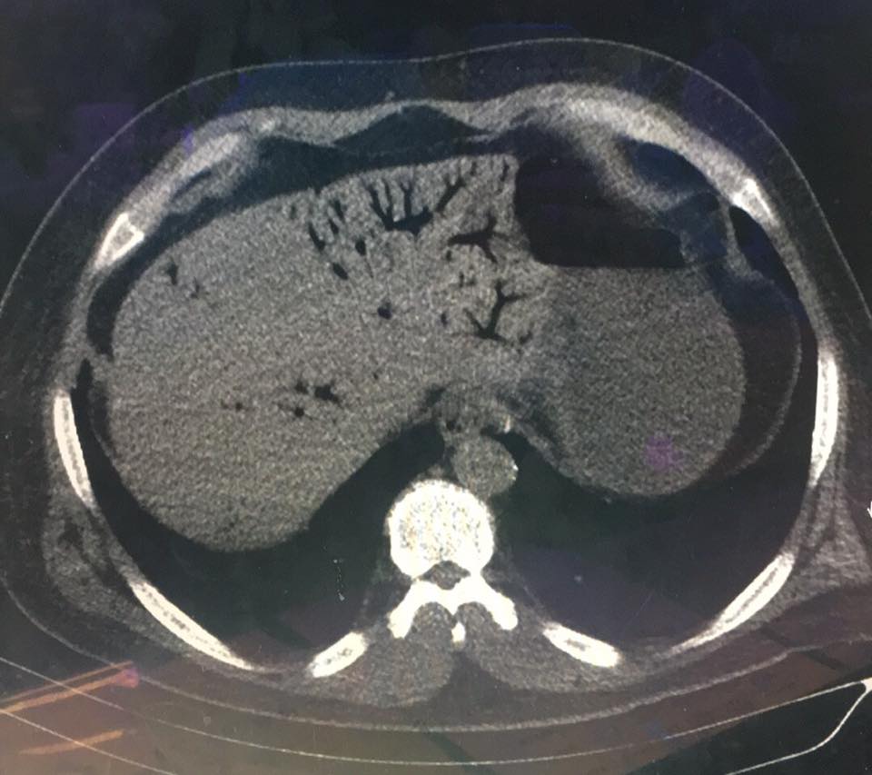
Pulmonary barotrauma 肺部氣壓傷. 也叫做 pulmonary overinflation 肺部過度充氣症, burst lung syndrome 肺爆裂症
上升過程因故意閉氣,咳嗽,嘔吐等動作造成聲帶關閉, 造成肺部氣壓大於外界, 通常發生於恐慌的潛水員或氣瓶用盡而快速上升,造成肺部過度膨脹引起氣壓傷, 在淺水也可能發生(例如游泳池) ,
肺部氣壓傷包括以下幾種
縱膈腔氣腫: 胸部x光可能沒有明顯異常. 通常不需要高壓氧治療, 症狀治療即可(疼痛則給予止痛), 氣體可能會沿著皮下跑到頸部引起頸部皮下氣腫, 肺部過度膨脹可能造成氣胸 (有些患者可能須用空針抽出氣胸部位的氣體, 有些可能需放置胸管)
如果氣體進入肺部靜脈循環, 可能之後進入動脈循環引起動脈氣栓, 對於氣栓最敏感的器官是腦部, 這種情況稱腦部動脈氣栓 cerebral arterial gas embolism , 但氣栓可能同時散步到其他器官或組織, 上升過程的氣壓傷害如果同時合併中樞神經受損症狀, 要考慮腦部動脈氣栓
有些人因為其他疾病, 可能在沒有閉氣上升, 沒有快速上升的情況出現肺部氣壓傷(例如肺囊腫 lungcyst, 肺部阻塞性疾病, 或其他可能造成肺臟局部氣體侷限的疾病)
closed glottis (holds breath, coughs, vomits)
most frequently seen in a rapid, panicked, out-of-air ascent
the expanding air may cause parenchymal lung injury.
This can occur even in shallow water (e.g., a swimming pool)
Also called pulmonary overinflation, burst lung syndrome
Pneumomediastinum
Symptomatic treatment, NO RECOMPRESSION.
CXR may subtle
Neck SubQ emphysema
Cervical spine x-ray Emphysema.
Pneumothorax
Aspiration of air
tube thoracostomy.
PULMONARY BAROTRAUMA
Air also expands within the lungs with ascent. If a diver breathing compressed air ascends with a closed glottis (holds breath, coughs, vomits), most frequently seen in a rapid, panicked, out-of-air ascent, the expanding air may cause parenchymal lung injury. This can occur even in shallow water (e.g., a swimming pool). Pulmonary barotrauma, also called pulmonary overinflation or burst lung syndrome, can lead to pneumomediastinum. This generally only requires symptomatic treatment and may be subtle on the chest radiograph.3 Mediastinal air can track superiorly into the neck, resulting in subcutaneous air on physical examination or
air on a cervical spine radiograph. Pulmonary overinflation injury can cause pneumothorax, requiring aspiration or air or tube thoracostomy. If air enters the pulmonary venous circulation, embolization of the gas through the arterial system occurs. The most sensitive end-organ to such embolization is the brain, and cerebral arterial gas embolism is the term applied to this condition, although the air emboli distribute to other tissues and organs.4 Any neurologic symptom or sign referable to the circulation to the CNS in the setting of barotrauma associated with ascent should be considered to be secondary to cerebral arterial gas embolism. The symptoms, signs, and treatment are discussed below in the section “Arterial Gas Embolism.” Pulmonary barotrauma (Figure 214-1) can occur without a rapid ascent or closed glottis in divers with congenital cysts, obstructive pulmonary disease, or other processes that cause air trapping.
其他上升過程的氣壓傷包括牙齒內氣泡膨脹, 會造成牙痛, 甚至牙齒填充物脫離,牙齒斷裂. 在潛水過程吞入胃部的氣體, 上升過程膨脹之後會造成腹部膨脹和絞痛.
OTHER BAROTRAUMAS OF ASCENT
An air pocket underneath a tooth may equilibrate with ambient pressure while diving, only to expand during ascent. This produces severe pain and may dislodge a filling or fracture a tooth. Swallowed air during diving may expand during ascent, rarely producing gastric distention and abdominal cramps.
DECOMPRESSION SICKNESS
減壓症的病理生理機轉, 與惰性氣體(主要是指氮氣)造成循環阻塞, 及組織發炎反應有關, 減壓症可能發生於使用壓縮氣體的潛水員, 涵箱工作人員, 高空飛行員, 太空人. 溶解入血漿的惰性氣體在壓力減小的時候會形成氣泡釋出, 不同組織對於惰性氣體有不同吸收率, 美國海軍發表了潛水計畫表, 提供不同條件的潛水免減壓極限, 其他海軍潛水計畫表提供各種長時間潛水的減壓程序, 使用數學模式計算免減壓時間的潛水電腦錶讓潛水員能安全潛水. 如果遵循潛水計畫表或潛水電腦錶不做極限潛水, 不太可能發生減壓症. 當然, 遵循安全規則也不能完全保證不出現減壓症.
要造成減壓症需要有足量的氣泡, 但有氣泡產生未必會形成減壓症, 需達到一定的閾值, 在連續多次潛水之後產生氣泡並不一定會造成減壓症. 顯然氣泡負載 bubble load 需要到達一定數值才會造成症狀, 氣泡產生的真實機制不明, 但先前存在於循環之中的氣泡微粒 gas micromnuclei 可能會累積形成氣泡窩 nidus, 這個推斷是基於新生氣泡所需的能量, 遠大於組織中惰性氣體達到飽和狀態所需能量(氣泡無中生有比較不容易). 氣泡可能直接在組織或血液循環中(通常是壓力較低的靜脈循環)產生, 傳統認為氣泡直接阻斷血液循環造成缺血, 但氣泡與血液-內皮細胞介面會啟動各種發炎反應及血栓生成, 活化上皮細胞, 導致中性球黏著與活化, 改變上皮細胞通透性, 導致液體流向細胞間隙. 此外, 減壓過程的刺激導致微粒 microparticle (雙層脂質膜微粒)於血管上皮細胞或其他細胞產生
在動物實驗直接注射這種 microparticles 會誘發與減壓症相同的臨床症狀
The pathophysiology of decompression sickness is related to the obstructive and inflammatory effects of inert gas bubbles in tissues and the vascular system.4 Decompression sickness may occur in divers breathing compressed air, caisson workers, high-altitude pilots, or astronauts. Bubbles may form when a body with additional inert gas in solution experiences a decrease in ambient pressure that causes liberation of the gas. Uptake of inert gas occurs at different rates in different tissues. The U.S. Navy publishes dive tables to provide the limits to a dive (measured by bottom depth and time) that can be undertaken without a decompression stop (“no decompression” or “no stop” dives). Other Navy tables provide a variety of decompression schedules for longer dives. A multitude of dive computers, often using proprietary mathematical models, provide divers with relatively safe diving limits. Decompression sickness is unlikely to occur if the limits of the dive tables or dive computer are followed, but compliance with dive table limits or a dive computer does not completely eliminate risk. Bubbles are necessary but not sufficient by themselves to cause
decompression sickness; bubbling occurs after many dive profiles that do not lead to decompression sickness. Obviously, there must be a threshold at which the bubble load causes symptoms. The exact mechanism of bubble formation is not known, although preexisting gas micronuclei in the circulation likely form a nidus for gas accumulation. This is inferred, because the energy required to form bubbles de novo is much higher than the energy state caused by the saturation of inert gas in tissue. 5 Bubbles may form directly in tissues or the circulation (usually the low-pressure venous circulation). Classically, it is thought that bubbles directly obstruct blood flow, leading to direct ischemia. Also, the air– blood and air–endothelial interfaces initiate a variety of inflammatory
and thrombotic processes; activate the endothelium, leading to neutrophil adhesion and activation; and change the permeability of the endothelium, resulting in third spacing of fluid. In addition, decompression stress induces the production of microparticles, which are lipid bilayer– enclosed membranous vesicles extruded from vascular endothelial and other cells. Injection of these microparticles in animal models creates a clinical condition consistent with decompression sickness.6 There are no current definitive diagnostic criteria for decompression sickness. The San Diego Diving and Hyperbaric Organizations criteria use a point system to identify dive injuries resulting in decompression sickness with a high degree of specificity.7 This is helpful to create databases of divers with decompression sickness to study outcomes and allow study of adjunctive therapies. Unfortunately, this system has relatively low sensitivity. Studies of therapies for decompression sickness often lack an acceptable case definition of decompression sickness.
CLINICAL FEATURES 減壓症臨床症狀
The most commonly used classification divides decompression sickness into two (or sometimes three) main groups (Table 214-2). We focus on type I and II for clarity. Type I is also called “pain-only” decompression sickness and involves the joints, extremities, and skin (“cutis marmorata”). Lymphatic obstruction can occur in type I, causing lymphedema, which usually takes days to resolve despite recompression therapy. Type II involves the CNS (mainly the spinal cord in compressed air divers and the brain in high-altitude decompressions), vestibular symptoms (“staggers”), and cardiopulmonary symptoms (“chokes”). To further complicate the
nomenclature and classification of decompression sickness, it can also occur when an arterial gas embolism (see below) causes inert gas to come out of solution after a dive profile that would otherwise not be expected to cause decompression sickness (called type III).8 Some advocate the use of the alternate term decompression illness, instead of differentiating between decompression sickness and cerebral arterial gas embolism, to encompass all pathologic syndromes following a reduction in ambient pressure.1
Generally, the symptoms of decompression sickness occur minutes to several hours after surfacing, but in rare cases, symptoms can occurdays after diving. Symptoms occurring between dives may improve during a subsequent dive (as recompression has occurred) but get worse upon resurfacing (as the inert gas load has increased and ambient pressure has decreased). Flying with the resultant decrease in ambient pressure may precipitate or worsen symptoms. For this reason, divers are generally advised to refrain from flying for at least 12 to 24 hours after the last dive depending on the nature of the diving exposure.9
Pain Divers with type I decompression sickness typically describe a deep pain, unrelieved but not worsened with movement. This pain can be attributed to or confused with pain caused by injury, potentially making accurate diagnosis difficult. Pain is thought to be due to distention from bubbles in ligaments or fascia, intramedullary bubbles at the ends of long bones, or the activation of stretch receptors caused by bubbles in tendons. The mechanism of simple distention of tissues is supported by the rapid improvement of symptoms with recompression. Common pain locations are knees and shoulders, and most often, only a single joint is involved. Decompression sickness in commercial and military divers, caisson workers, and aviators tends to manifest most often as joint pain. Sport divers, who usually perform multiple dives, often over a period of days, are more prone to spinal cord effects. Poorly localized and difficult-to-describe back or abdominal pain may herald the more serious signs of spinal cord involvement. Pulmonary Symptoms Pulmonary symptoms, generally seen usually only after more prolonged exposures, are caused by large numbers of pulmonary artery bubbles and include symptoms of cough, hemoptysis, dyspnea, and substernal chest pain. Cardiovascular collapse can occur. Neurologic Symptoms The classic description of divers with neurologic decompression sickness (type II) can begin with a sensation of truncal constriction or girdle-like pain. Often a wooly feeling begins in the feet, developing into an ascending paralysis, producing symptoms of transverse myelitis. This form is usually rapid in onset and has a tendency to affect the lower cervical and thoracic regions. However, in type II decompression sickness, neurologic deficits do not necessarily cause distinct spinal cord syndromes (i.e., an anterior or posterior spinal artery syndrome), nor will a definitive level necessarily be found, as lesions may be scattered throughout the spinal cord.10 Autonomic involvement, with resulting incontinence and sexual dysfunction, is not uncommon. The pathophysiology of spinal cord decompression sickness seems to be initial bubbling in the low-pressure venous plexus system that first impedes and thenobstructs venous outflow from the cord. Decreasing venous blood flow prevents dissolved nitrogen in spinal cord tissues from egressing, and in situ bubbles within the spinal cord develop (called autochthonous bubbles).
Vestibular Symptoms Vestibular decompression sickness usually occurs after deep, long dives, although it has been reported in sport divers. Signs are vertigo, hearing loss, tinnitus, and disequilibrium. The vestibular syndrome can be differentiated from inner ear barotrauma mainly by the history, because patients with inner ear barotrauma develop symptoms in the water and, generally, immediately after a forced Valsalva maneuver to equalize the middle ear pressure.11 Other, nonspecific symptoms such as headache, nausea, dizziness, or unusual fatigue are also reported. It may be difficult to differentiate fatigue from decompression sickness from the expected fatigue from the exertion of diving.
Patent Foramen Ovale
The association between decompression sickness and patent foramen ovale is unclear. There appears to be an increased prevalence in patients with inner ear and cutaneous decompression sickness. It is reasonable to screen divers with recurrent, unexplained decompression sickness for a patent foramen ovale. Closure of a large defect will reduce arterialization of venous gas emboli, although it has yet to be shown if such closure will reduce the incidence of subsequent decompression sickness.12
ARTERIAL GAS EMBOLISM
Arterial gas embolism occurs when air enters the left side of the vascular system. In the setting of diving, this most often results from pulmonary barotrauma. Arterial gas embolism can also occur as a complication of certain medical procedures, such as central vascular catheterization and cardiac bypass. Air inadvertently introduced into the venous circulation can cross from the right side of the circulation from intracardiac or pulmonary arteriovenous shunts. Air bubbles may also arterialize through these same shunts, sometimes making the source of arterial bubbles difficult to determine.13 Whatever the source, when air embolizes systemically, distribution depends mainly on blood flow and not gravity. Clinical Features The most dramatic effect of arterial gas embolism is
on the brain, resulting in a variety of stroke syndromes, symptoms, and signs, depending on the part of the brain affected. Rarely, diving-related arterial gas embolism from pulmonary barotrauma causes immediate apnea and cardiac arrest. The mechanism of cardiovascular collapseappears to be air in the entirety of the large arteries and veins of the central vascular bed.14 The effects of arterial gas embolism secondary to pulmonary barotrauma usually occur on ascent or immediately upon surfacing. If the victim does not die immediately, the symptoms of cerebral arterial gas embolism often include loss of consciousness, seizure, blindness, disorientation, or hemiplegia. Symptoms may spontaneously improve as the gas enters the venous cerebral circulation after a spike in blood pressure. Sometimes, by the time the patient reaches the clinician, the only signs that remain are subtle defects. In particular, parietal lobe signs and symptoms are easily overlooked. A cascade of inflammatory processes also occurs in air embolism, just as in decompression sickness.4 Laboratory Testing The hematocrit may elevate from hemoconcentration and third spacing of fluids. The creatine phosphokinase (and other enzymes such as lactate dehydrogenase, alanine aminotransferase,
and aspartate aminotransferase) will become elevated secondary to the systemic distribution of bubbles. The degree of elevation of creatine phosphokinase corresponds to the embolism severity. Cardiac troponins may also be elevated and most likely do not represent occlusive coronary artery disease.4
Treatment of Decompression Sickness and Arterial Gas Embolism
The treatment includes administering 100% oxygen, increasing tissue perfusion with IV fluids, and rapid recompression. Some advocate placing patients with air embolism in the Trendelenburg position or in the left lateral decubitus position to “trap” air in the left ventricle. By the time the victim is brought onto the dive boat or the ambulance arrives, the air has usually been distributed, and the Trendelenburg position merely increases intracranial pressure, decreases cerebral perfusion, and interferes with other first aid measures. Nonetheless, some divers with arterial gas embolism have collapsed when placed in a sitting or standing position. As a result, a supine position—not Trendelenburg position—is recommended for patients with arterial gas embolism. Vomiting patients
should be placed in the lateral decubitus position to prevent aspiration. Recompression therapy with hyperbaric oxygen treats by several mechanisms. See the chapter 21, titled “Hyperbaric Oxygen Therapy” for detailed discussion. The administered pressure decreases the size of bubbles, and the high partial pressure of oxygen in solution increases inert gas washout from bubbles and tissue. Mass action dictates a gas will travel down pressure gradients; therefore, nitrogen will move from bubbles with a high partial pressure of nitrogen into plasma, where it will travel to the lungs and be exhaled. Conversely, oxygen from plasma with a high partial pressure of oxygen will enter bubbles, but ultimately will diffuse into cells and be metabolized, further reducing bubble size. Hyperbaric oxygen also decreases tissue edema, increases oxygen delivery to ischemic tissues, and reduces neutrophil adhesion to the endothelium and neutrophil activation.15
Recompression using the U.S. Navy Treatment Table 6 is a commonly used method of management for decompression sickness, employing a maximal treatment pressure of 2.8 ATA (60 fsw). Table 6 is also used for air embolism, although some advocate an initial pressurization to 6 ATA (165 fsw) to maximize bubble compression, then continuation at 2.8 ATA (U.S. Navy Table 6A). Different treatment tables are used in other parts of the world, and there is some experience using lower treatment pressures for decompression sickness in monoplace chambers with reportedly comparable results.16 Some patients may benefit from repeated treatments if symptoms do not fully resolve. Recompression should occur as soon as possible, and it should not be withheld in cases
with delayed presentation.4 Additionally, for divers who have missed needed decompression stops because of an emergency ascent or nonadherence to appropriate diving tables, it may be appropriate for them to undergo recompression therapy even if asymptomatic. U.S. Navy Table 5 recompression would usually be adequate in such a circumstance. The administration of IV lidocaine as a therapeutic adjunct for cerebral arterial gas embolism has been advocated, because it appears to decrease neuropsychiatric deficits when given during anesthesia for cardiac procedures requiring bypass,17-19 since bypass operations commonly cause the entry of air into the arterial system. Dosing of lidocaine in this setting is not standardized, although typical cardiac dosing is commonly used.20 The Divers Alert Network (telephone: 1-919-684-9111; Web site: http://www.diversalertnetwork.org) has staff available 24 hours a day to provide assistance to divers and to help clinicians treat patients with decompression sickness or arterial gas embolism. The Divers Alert Network can provide information and the location of the nearest recompression facility around the world.
SPECIAL CONSIDERATIONS
ASTHMA
There is some debate over the safety of diving for individuals with asthma. Although the relative risk of pulmonary barotrauma may be higher in asthmatics (possibly as much as twice that of the general diving population), the absolute risk is still low because of the rarity of pulmonary barotrauma in diving (approximately 1 in 125,000 dives).21 A physician who specializes in diving medicine should examine divers or potential divers with asthma, and an exercise pulmonary function test should be performed. Asthmatics can be cleared for diving if, using their usual medications, they have a normal exercise pulmonary function test and if they understand the potential increased risk of pulmonary barotrauma. 22 A diver who develops a lung injury that cannot be explained
by the circumstances of the dive (i.e., the diver did not have a rapid, breath-holding ascent) should be evaluated for congenital or acquired structural lung disease and should probably no longer dive.
IMMERSION PULMONARY EDEMA
Pulmonary edema can occur while diving. Because the first reported cases occurred in cold water, this condition was first described as “cold water” or “cold-induced” pulmonary edema. However, many cases have subsequently been reported in warm water, up to 27°C (80.6°F).23 Typical symptoms of pulmonary edema (dyspnea, chest discomfort, coughing up pink frothy secretions) occur at depth and usually improve over time or with standard treatments for pulmonary edema. The cause is unknown despite human studies.24 This syndrome generally occurs in divers with no structural or ischemic heart disease, but diagnostic evaluation for heart conditions is indicated for those with risk factors for underlying heart disease. Immersion pulmonary edema is not caused by decompression and is not treated with recompression therapy. Interestingly, some divers will experience repeated episodes, whereas others may never experience another episode.
NITROGEN NARCOSIS
Inert gas narcosis occurs when air is breathed at a depth of 100 fsw or greater. Symptoms include loss of fine motor skills and high-order mental processes as well as behavior similar to that seen in alcohol intoxication. Symptoms increase as depth is increased beyond 100 fsw. Divers have codified this increase in symptoms as the “martini rule” (with many variations). A common description is that each 33 ft of depth (1 ATA) in excess of 100 fsw is the equivalent to drinking one martini. Nitrogen narcosis can cause divers to engage in dangerous or foolish activities during deep dives. At depths greater than 300 fsw, unconsciousness may occur from the anesthetic effect of nitrogen. At depths greater than 200 fsw, helium is often used in place of nitrogen in gas mixtures to prevent
nitrogen narcosis.
OXYGEN TOXICITY
Oxygen toxicity usually affects the lungs or brain, depending on the partial pressure of oxygen delivered and duration of exposure. Pulmonary oxygen toxicity generally occurs at lower partial pressures of oxygen but with longer exposures, whereas cerebral oxygen toxicity occurs at high partial pressures with generally short exposures. Pulmonary oxygen toxicity can occur at partial pressures of oxygen at or below 1 ATA, for example, in patients requiring prolonged mechanical ventilation with high fractions of inspired oxygen. Pulmonary oxygen toxicity is unusual in diving.
Cerebral oxygen toxicity most often occurs with partial pressures of oxygen >1.4 ATA in the water. Some divers may breathe “nitrox” or oxygen-enriched air with fractions of oxygen of 32% to 36%. Therefore, cerebral oxygen toxicity can occur at lesser depths and actually is the factor that limits diving depth with nitrox. Additionally, there are rebreather systems (closed-circuit systems), with the diver breathing within a continuous circuit of gas that has a very high fraction of oxygen (>95%), with carbon dioxide being scrubbed out. With these systems, cerebral oxygen toxicity can occur at depths as little as 25 ft. Signs and symptoms of cerebral oxygen toxicity include twitching, nausea, paresthesias, dizziness, and seizures. If a seizure develops in the water as the initial manifestation of cerebral oxygen toxicity, drowning may result. High partial pressures of oxygen are used clinically in hyperbaric chambers (2.4 ATA, 2.8 ATA, and sometimes 3.0 ATA), but cerebral oxygen toxicity in this setting is rare, reported in <1 per 1000 patients.
This is because patients in hyperbaric chambers are dry, warm, and at rest, while divers are wet, often cold, and exerting themselves—and all of these latter factors exacerbate cerebral oxygen toxicity. Cerebral oxygen toxicity is affected by partial pressure of arterial carbon dioxide and cerebral blood flow and may be caused by an increase in nitric oxide production, although this is still an area of active investigation.25 Besides nitrogen narcosis and oxygen toxicity, other gas-related conditions important in diving medicine are toxicity from carbon monoxide and the adverse effects of elevated partial pressures of carbon dioxide. Additional issues, especially with very deep dives, include heat loss from breathing helium and the high-pressure nervous syndrome, characterized by tremor and loss of fine motor function caused by the direct effects of pressure.
OTHER CONDITIONS
在一般人會出現的疾病, 壓縮空氣潛水同樣可以發生, 所以不要忘了其他常見的疾病,例如心肌梗塞,肺栓塞,中風,癲癇,腦炎,甚至闌尾炎, 另外,潛水意外或突發性心跳停止可能會被誤認為溺水.
Injuries and medical conditions occurring during and immediately after compressed air diving are often misattributed as decompression sickness or cerebral arterial gas embolism. Be aware that any medical condition can occur under the water. Acute myocardial infarction, pulmonary embolism, stroke, seizure, encephalitis, and even appendicitis have been erroneously attributed to diving. True diving accidents or cardiac sudden death can be misattributed to drowning—a common final pathway of submersion.26
Tintinalli 建議可以諮詢 DAN 網站.
https://www.diversalertnetwork.org/medical/faq/
PFO 軟圓孔閉合不全 大約 25-30% 的盛行率, 但多數人的孔很小, 不足以造成症狀, 且左心房的壓力比右心房大, 血液不太會從右心房流向左心房
某些人的PFO可能比較嚴重, 造成右心房的氣泡直接流入左心房, 進入全身循環, 引起嚴重的氣栓
如果在正常的潛水條件下發生嚴重減壓病, 需請醫師評估是否存在PFO. 但多數潛水醫學醫師認為, PFO造成的危害不大, 不需要將篩檢所有潛水員
In approximately 25-30 percent of individuals, this communication persists as a small opening, called a patent foramen ovale (PFO).
At present, most diving physicians agree that the risk of a problem associated with a PFO is not significant enough to warrant widespread screening of all divers. An episode of severe decompression illness that is not explained by the dive profile should initiate an evaluation for the existence of a PFO.
https://www.diversalertnetwork.org/medical/faq/Patent_Foramen_Ovale_PFO
美國海軍潛水操作手冊 17-8 開始介紹 DECOMPRESSION SICKNESS
http://www.navsea.navy.mil/Portals/103/Documents/SUPSALV/Diving/US%20DIVING%20MANUAL_REV7.pdf?ver=2017-01-11-102354-393
減壓症發作時間, 42%在一小時內發生, 98%在 24 小時內發生
•U.S. Navy Treatment Table 6
–maximal treatment pressure of 2.8 ATA (60 fsw).
–also used for air embolism
–some advocate an initial pressurization to 6 ATA (165 fsw) to maximize bubble compression, then continuation at 2.8 ATA (U.S. Navy Table 6A).
•Additionally, for divers who have missed needed decompression stops because of an emergency ascent or nonadherence to appropriate diving tables, it may be appropriate for them to undergo recompression therapy even if asymptomatic.
•U.S. Navy Table 5 recompression would usually be adequate in such a circumstance.
US NAVY TABLE 5


下面的連結應該是給潛水的一般民眾看得網誌. 介紹各種潛水造成的疾病(外傷)
https://www.diversalertnetwork.org/health/ears/perforated-eardrum-tympanic-membrane-rupture
減壓病
惰性氣體在壓力上升時會溶解進入血中, 當壓力減小時釋放出來,. 形成小氣泡, 如果壓力減少太快, 氣泡產生過快, 會造成減壓病, 治療方式需 HBO 高壓氧
HBO 能加速惰性氣體釋出體外, 如果沒有 HBO, 症狀輕微持續超過 24 小時的病患, 神經功能如果正常, 可使用氧氣治療
如果病患須轉送到高壓氧中心, 飛機需艙內加壓, 維持在海平面一大氣壓的壓力, 如果無法加壓, 建議飛行在最低的高度, 例如直升機運送時.
HBO 設定多數是 2.8 atm 持續 60-90 分鐘, 之後逐步減壓, 遵循美國海軍潛水操作治療表.
多數病患經過單次治療會有反應, 氣泡在體內可持續數天, 所以即使耽擱很多天, HBO 仍可能有效果.
DECOMPRESSION SICKNESS 減壓病
Decompression sickness is due to the formation of nitrogen bubbles in body tissue and circulation during decompression (see chapter 214, “Diving Disorders”).7 During pressurization, inert gas (nitrogen if breathing air) is dissolved in body fluids. Upon reduction in ambient pressure, the dissolved gas comes out of solution and forms small bubbles in the tissue and circulation. HBO is an effective treatment because the increase in ambient pressure reduces gas bubble volume and the supplemental oxygen hastens inert gas diffusion out of the body. Decompression sickness should be treated with HBO when such therapy is available and no contraindication exists. If HBO therapy is not available, patients with mild symptoms and neurologic stability for longer than 24 hours may be treated with supplemental oxygen therapy alone. If a patient must be transported by air to a hyperbaric facility, use pressurized aircraft to maintain sea-level pressure or transport at the lowest possible altitude in a nonpressurized craft, such a helicopter. A variety of HBO regimens are used to treat decompression sickness,8 but most have in common pressurization to 2.8 ATA for 60 to 90 minutes, followed by stepwise decompression, similar to U.S. Navy Diving Manual Treatment Table 6. Most patients respond to a single treatment. Gas bubbles can persist for several days, and HBO may be beneficial even when begun after long delays.8


沒有留言:
張貼留言